
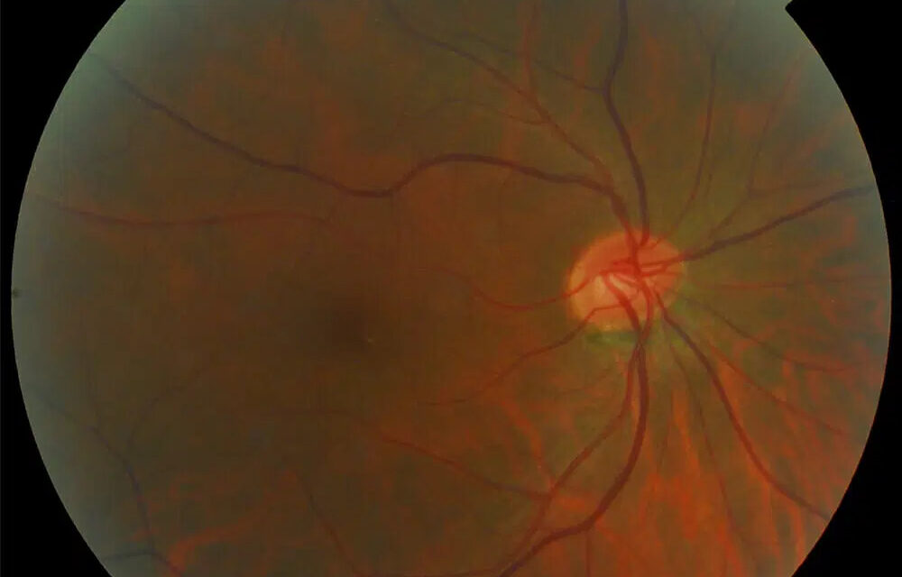
Toxic and nutritional optic neuropathy describes a group of conditions producing bilateral, symmetric, progressive optic nerve dysfunction from either direct toxic injury to the optic nerve or from deficiency of nutrients the nerve needs to function normally.
The two categories are grouped together because they share a strikingly similar clinical presentation and, in many real-world cases, actually overlap in the same patient.
Chronic alcohol use, for example, often combines direct toxic exposure with the nutritional deficiency that frequently accompanies heavy drinking, making it genuinely difficult in practice to attribute a given patient’s optic neuropathy to one mechanism alone.
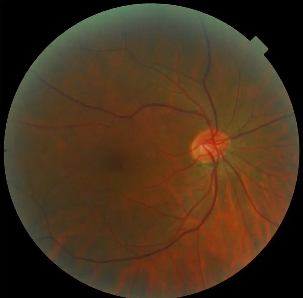
Why This Pattern Is Distinctive
Unlike most other optic neuropathies discussed elsewhere on this site, toxic and nutritional optic neuropathy is characteristically bilateral and symmetric from the outset, and develops gradually rather than acutely.
It preferentially affects the papillomacular bundle — the tightly packed group of fibers running from the fovea directly to the optic disc — producing a central or cecocentral scotoma rather than the more common patterns of visual field loss seen in other optic nerve diseases.
This specific combination of bilateral, symmetric, gradual, central field loss is the pattern that should prompt consideration of a toxic or nutritional cause before other, more common optic neuropathies.
Recognizing this pattern early has real practical value, since it directs the workup toward a detailed history and targeted laboratory testing rather than the more extensive, and often less immediately productive, imaging-focused workup pursued for other optic neuropathy presentations.
Toxic Causes
- Ethambutol — a well-known, dose-dependent cause requiring baseline and periodic visual monitoring in any patient started on this antituberculous medication
- Methanol — classically from ingestion of contaminated or counterfeit alcohol, capable of producing severe, often irreversible bilateral vision loss
- Amiodarone
- Chloramphenicol, particularly with prolonged use
- Chronic tobacco and alcohol use — historically described together as “tobacco-alcohol amblyopia,” now generally understood to reflect a combination of direct toxicity and the nutritional deficiency (particularly B vitamins) that often accompanies heavy, chronic use of both substances
- Lead and other heavy metal exposure
Fundus Explorer Pro
Photograph the retinal findings described here with the phone already in your pocket — 22 D optics and built-in illumination in one handheld unit.
From Choroida — the team behind this siteNutritional Causes
- Vitamin B12 deficiency
- Folate deficiency
- Thiamine (B1) deficiency
- Copper deficiency, an increasingly recognized cause, particularly after bariatric surgery where malabsorption of trace minerals can be overlooked relative to more commonly screened-for deficiencies
- General malnutrition or malabsorption from any cause, including inflammatory bowel disease, prior gastric surgery, or restrictive diets
Clinical Presentation
Patients describe gradual, painless, bilateral blurring of central vision, often first noticed with reading or other detailed visual tasks, evolving over weeks to months rather than the acute or subacute onset typical of optic neuritis or ischemic optic neuropathy.
Color vision, specifically red-green discrimination, is often affected early and disproportionately relative to visual acuity, and can be a useful, sensitive marker of subclinical involvement even before acuity itself has dropped significantly, making routine color vision testing a worthwhile addition when this diagnosis is being considered or monitored.
Exam Findings
- Reduced visual acuity, bilateral and roughly symmetric
- Central or cecocentral scotoma on visual field testing
- Impaired color vision, often out of proportion to visual acuity loss in earlier stages
- The optic disc can appear entirely normal early in the course, which is an important and sometimes counterintuitive point — normal-appearing discs do not exclude toxic or nutritional optic neuropathy
- Temporal disc pallor developing later, as axonal loss accumulates, reflecting the preferential involvement of the papillomacular bundle
Diagnostic Evaluation
A detailed history is the single most valuable diagnostic tool: medication list (with specific attention to ethambutol and amiodarone), alcohol and tobacco use, dietary history, any prior bariatric or gastrointestinal surgery, and possible toxic exposures.
Laboratory testing for B12, folate, and, where indicated, copper and thiamine levels helps identify a treatable nutritional cause.
Neuroimaging is appropriate to exclude a compressive lesion when the presentation is atypical or when initial workup does not identify a clear toxic or nutritional explanation, because compressive optic neuropathy can occasionally mimic this pattern.
Genetic testing for hereditary optic neuropathies, particularly Leber hereditary optic neuropathy and dominant optic atrophy, is also worth considering in cases without a clear toxic, nutritional, or compressive explanation, since these inherited conditions can produce a similar bilateral, symmetric, central visual field pattern.
Management
Removing or discontinuing the offending toxic agent — stopping ethambutol, addressing alcohol use, correcting the source of heavy metal exposure — is the essential first step, and for many toxic causes, the degree of recovery depends heavily on how promptly the exposure is stopped once symptoms begin.
Nutritional causes are treated with targeted replacement (vitamin B12 injections, folate supplementation, copper repletion), often producing meaningful visual recovery over weeks to months once the deficiency is corrected, when treatment begins before extensive axonal loss has occurred.
Because delayed recognition allows more irreversible axonal damage to accumulate, both toxic and nutritional optic neuropathy reward early diagnosis disproportionately.
The same underlying process caught early is often substantially reversible, while advanced, longstanding disease with established optic atrophy typically is not, regardless of how thoroughly the underlying cause is subsequently addressed.
This makes patient and, where relevant, prescriber education a genuinely important preventive measure — for example, ensuring that patients on ethambutol understand why they need scheduled visual monitoring, rather than only presenting once symptoms have already developed.


Document what you see
Two smartphone imaging tools built for everyday clinic use — one for the slit lamp, one for the fundus.
From Choroida — the team behind this siteReferences
- Grzybowski A, Zülsdorff M, Wilhelm H, Tonagel F. Toxic optic neuropathies: an updated review. Acta Ophthalmologica.
- Sharma P, Sharma R. Toxic optic neuropathy. Indian Journal of Ophthalmology.
- American Academy of Ophthalmology. Basic and Clinical Science Course, Section 5: Neuro-Ophthalmology.
