
CASE REPORT
A 15-year-old, healthy female patient was first noted to have a retinal abnormality during a routine visit to her optician.

She was asymptomatic and had no significant ophthalmic or medical history or any history of medication.
Visual acuities were counting fingers at 1 meter in the right eye and 6/5, N5 in the left eye. Slit lamp biomicroscopy of the right eye revealed a normal vitreous cavity unaccompanied by signs of active uveitis.
The striking abnormality was the presence of a subfoveal grayish lesion at the level of the retinal pigment epithelium (RPE) measuring approximately 1/3 disc diameter in size surrounded by subretinal exudates associated with a fluffy white appearance.
There was evidence of neurosensory retinal detachment overlying and surrounding the entire lesion as noted by the tenting up of the retina in the macular region.
Both optic discs were pink and healthy. Fluorescein angiography demonstrated hyper-fluorescence of a subfoveal lesion. The clinical picture was thought to be consistent with atypical Best’s disease.
However, the electrooculogram (EOG) and Ganzfeld Electroretinogram (ERG) investigations, performed to the international standards, were normal, although, in the right eye, the five main responses of the ERG were slightly reduced in comparison to the left.
The findings were not different from the photopic and scotopic ERG. In contrast, the Wide Field Multifocal Electroretinogram (mfERG) showed a marked reduction in the central responses in the right eye.
Best’s disease and diffuse retinal dystrophy were therefore excluded. A revised diagnosis of unilateral acute idiopathic maculopathy was made.
Acute Idiopathic Maculopathy DISEASE entity
Acute Idiopathic Maculopathy is an uncommon disease of the retinal pigment epithelium that affects young adults. It presents as a sudden, severe unilateral central vision loss most commonly following a flu-like illness.
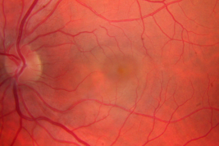
Unilateral Acute Idiopathic Maculopathy (UAIM) was first described as a syndrome by Yannuzzi, et al in 1991.
His report consisted of nine young patients who developed significant unilateral vision loss (20/200 or worse) associated with a characteristic exudative detachment of the macula with grayish thickening of the underlying retinal pigment epithelium (RPE).
Patients experienced spontaneous resolution of their symptoms over the course of several days to weeks with near complete recovery of vision, a “bulls-eye” appearance in the macula persisted.
While his report discussed unilateral vision loss, bilateral disease has been reported as well leading some to propose renaming the disease Acute Idiopathic Maculopathy (AIM).
AIM by definition is not linked to an underlying disease process or known cause. It notably affects younger Caucasian individuals and it appears to split equally between sexes.
A preceding flu-like illness has been reported in several cases and elevated Coxsackie Virus titers were reported in a patient presenting with bilateral disease.
Clinical diagnosis
The diagnosis of this disease is clinical and supported by ancillary testing including Optical Coherence Tomography (OCT) and Fluorescein Angiography (FA).
Diagnostic procedures
In the acute phase of maculopathy spectral-domain OCT (SD-OCT) and STRATUSOCT show abnormal heterogeneous hyperreflective thickening at the level of the outer retina and RPE in the foveal region.
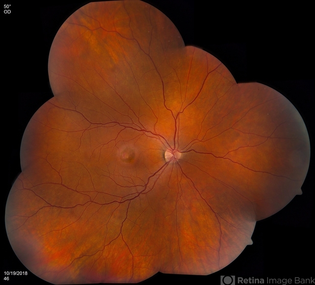
Hyporeflective exudation and subretinal fluid with detachment and disruption of the photoreceptor outer segment can also be seen.
Swept- Source Optical Coherence Tomography (SS-OCT) demonstrates similar findings. Upon resolution of the disease, the neurosensory detachment often resolves completely, however, patients may develop a focal submacular scar with persistent metamorphopsia. Choroidal neovascular membranes (CNV) can be seen as a complication of AIM.
A case report using Swept-Source Optical Coherence Tomography Angiography demonstrated early irregular hyperfluorescence and hypofluorescence at the level of the retinal pigment epithelial lesion, followed by complete staining of the overlying neurosensory retinal detachment in the late phase of the study.
The superficial and deep layers of the retinal capillary plexus were preserved and a dark pattern at the choriocapillaris segmentation line was observed when compared to the right eye.
Indocyanine green angiography shows a hypofluorescent lesion with some hyperfluorescence in the mid-late phase.
Differential diagnosis
The differential diagnosis of Acute Idiopathic Maculopathy includes diseases that present with a focal area of subretinal fluid (SRF) leading to profound central vision loss.
Central Serous Retinopathy, Harada Syndrome, and APMPEE are included in the differential and can generally be excluded by their FA findings.
The possible relationship between AIM and APMPPE has been suggested due to the overlap of many common features including the rapid onset and resolution of symptoms and the following of a prodromal flu-like illness.
Also, the various macular dystrophies should be considered, however, the progressive loss of vision and pigmentary changes associated with SRF can help distinguish them from AIM.
Fundus Explorer Pro
Photograph the retinal findings described here with the phone already in your pocket — 22 D optics and built-in illumination in one handheld unit.
From Choroida — the team behind this siteAcute Idiopathic Maculopathy MANAGEMENT
Historically, AIM has been misdiagnosed and interventions such as photodynamic therapy, systemic corticosteroids, and intravitreal triamcinolone have been prescribed.
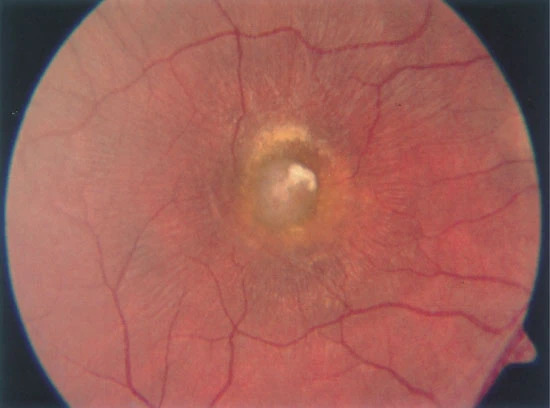
No treatment has been proven to work; it is a self-limiting condition. Patients presumed to have AIM should be observed clinically and with ancillary testing for spontaneous resolution. Viral serology may be obtained to identify an underlying cause.
Medical therapy:
No medical therapy is indicated as the maculopathy resolves on its own over the course of several days to weeks.
Medical follow-up:
Patients should be followed with serial fundus examination and OCT until resolution has occurred.
Prognosis
The overall prognosis for this disease is favorable as most patients achieve full visual recovery upon resolution of the maculopathy.


Document what you see
Two smartphone imaging tools built for everyday clinic use — one for the slit lamp, one for the fundus.
From Choroida — the team behind this siteREFERENCES
- “Unilateral Acute Idopathic Maculopathy.” Unilateral Acute Idopathic Maculopathy. https://retinavitreous.com/diseases/aim.php Retina Vitreous Associates of Florida, n.d. Web. 29 Nov. 2016.
- Fuente, Miguel A. De La, and Rubén Cuadrado. “Unilateral Acute Idiopathic Maculopathy: Angiography, Optical Coherence Tomography and Microperimetry Findings.” Journal of Ophthalmic Inflammation and Infection. Springer-Verlag, 24 Nov. 2010. Web. 26 Nov. 2016.
- Yannuzzi LA, Jampol LM, Rabb MF, Sorenson JA, Beyrer C, Wilcox LM., Jr Unilateral acute idiopathic maculopathy. Arch Ophthalmol. 1991;109:1411–1416. [PubMed]
- Ghazi,Nicola G., MD, Armand Daccache, MD, and Brian P. Conway, MD. “Acute Idiopathic Maculopathy.” AAO Journal. American Academy Of Ophthalmology, May 2007. Web. 26 Nov. 2016.
- “The Founder of VRMNY, Dr. Yanuzzi Has Made Numerous Innovative.” Vitreous Retina Macula Consultants of New York. VRMNY, n.d. Web. 29 Nov. 2016.
