
Parinaud oculoglandular syndrome is a unilateral granulomatous conjunctivitis accompanied by ipsilateral preauricular or submandibular lymph node enlargement.
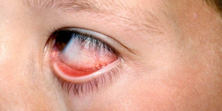
It is uncommon, but it is a classic pattern, and recognizing it points toward a short list of infectious and inflammatory causes.
The most frequent cause worldwide is cat scratch disease due to Bartonella henselae.
What Is Parinaud Oculoglandular Syndrome?
The syndrome combines three findings: a follicular or nodular granulomatous lesion of the conjunctiva, a tender regional node, and often a low-grade fever or malaise.
The conjunctival lesion is usually a red, elevated nodule or a patch of ulcerated, necrotic tissue, and it is often found in the tarsal conjunctiva or fornix.
The node is typically in the preauricular area, and it may suppurate.
The condition is distinct from Parinaud dorsal midbrain syndrome, which has the same eponym but affects vertical gaze and the pupils.
Causes
- Bartonella henselae (cat scratch disease): the most common cause, usually after a scratch or lick from a cat or kitten
- Tularemia: from handling infected rabbits or ticks, with a more severe systemic illness
- Tuberculosis and atypical mycobacteria: a chronic, indolent course
- Syphilis: primary or secondary lesions of the conjunctiva
- Sporotrichosis, lymphogranuloma venereum, and mumps: less common, but reported
- Lymphoma and sarcoidosis: noninfectious causes that need to be considered when the lesion persists
The history is the most efficient diagnostic tool, and animal exposure, travel, sexual history, and occupation should be asked about directly.
All-fit Slit-Lamp Adapter
Record and share exactly what you see at the slit lamp. One adapter fits any slit lamp or surgical microscope — and any smartphone.
From Choroida — the team behind this siteClinical Examination
Examine the conjunctiva with the lids everted, and look for a nodule, a membrane, or an ulcer.
Palpate the preauricular, submandibular, and cervical nodes, and record their size and tenderness.
Check the cornea and the posterior segment, because Bartonella can also cause neuroretinitis and other intraocular findings (see Bartonella henselae neuroretinitis).
A systemic review, including fever, weight loss, cough, and skin lesions, helps to find the cause.
Investigations
Serology for Bartonella is the first test in most cases, with PCR available on tissue or pus.
A conjunctival swab or biopsy for culture, PCR, and histology is reasonable when the lesion persists or the diagnosis is uncertain.
Tuberculosis testing and syphilis serology should be done when the history or the course fits.
Chest imaging is justified when sarcoidosis or tuberculosis is possible (see ocular tuberculosis).
Management
Cat scratch disease is usually self-limited and resolves over weeks to months.
Antibiotics such as azithromycin or doxycycline shorten the course in many patients, and they are used when the illness is significant or the patient is immunocompromised.
Suppurative nodes may need aspiration, and tularemia, tuberculosis, and syphilis need the specific regimen for each infection.
Immunocompromised patients are at risk of disseminated disease and should be referred to an infectious disease specialist.
Patients should be advised about avoiding scratches and licks from cats, and about washing any wound promptly.
Bartonella Infection in Detail
In cat scratch disease, a papule or pustule often appears at the site of inoculation a few days after contact, and the regional lymph nodes enlarge one to three weeks later.
When the inoculation site is the eye or the periocular skin, the conjunctival lesion is the primary site, and the preauricular node is the draining station.
Kittens are the usual source, and fleas appear to transmit the organism among cats, so the exposure may not involve a bite or a scratch that the patient remembers.
Most immunocompetent patients recover without sequelae, although the node may remain enlarged for weeks or months.
Clinical Pearls
A unilateral follicular conjunctivitis with a tender node that does not resolve in two weeks should prompt a more careful history and a swab.
A single node that is hard, fixed, and painless should not be assumed to be infectious, and lymphoma or metastasis may need to be excluded.
Documenting the size of the node and the conjunctival lesion with photographs helps to judge the response to treatment.
When to Refer
Refer when the diagnosis is uncertain after the first round of investigation, when the lesion is ulcerated or necrotic, or when the patient has systemic features such as weight loss or night sweats.
Immunocompromised patients, including those with HIV infection or on immunosuppressive treatment, are at risk of disseminated bartonellosis and need prompt assessment.
Any patient with a visual complaint should have a dilated fundus examination.
Warm compresses over the node and analgesia help symptoms, and the lesion is not contagious from person to person.
Patients with a persistent or enlarging node after treatment should have it reassessed, and an ultrasound or a fine-needle aspirate may be needed to separate infection from other causes.


Document what you see
Two smartphone imaging tools built for everyday clinic use — one for the slit lamp, one for the fundus.
From Choroida — the team behind this siteReferences
- Margileth AM, Wear DJ, English CK. Systemic cat scratch disease: report of 23 patients with prolonged or recurrent severe bacterial infection. J Infect Dis. 1987;155:390-402.
- Cunningham ET, Koehler JE. Ocular bartonellosis. Am J Ophthalmol. 2000;130:340-349.
- Salmon JF. Kanski’s Clinical Ophthalmology: A Systematic Approach. 9th ed. Elsevier; 2020.
- American Academy of Ophthalmology. Basic and Clinical Science Course, Section 9: Uveitis and Ocular Inflammation. AAO; current edition.

Writes and reviews the clinical articles on Educate. Every post is checked against peer-reviewed sources before it goes live.


