
When a gonioscopy lens shows a wavy, irregular band of brown pigment lying in front of Schwalbe line, the examiner has found a Sampaolesi line.
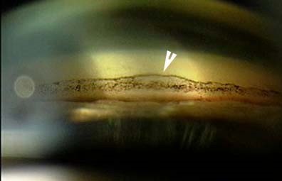
It is not a diagnosis by itself, but it should prompt a search for exfoliation material on the lens, and it has implications for glaucoma risk.
Where it lies
Schwalbe line marks the end of Descemet membrane and the front edge of the angle.
Normally, little or no pigment lies anterior to it.
In a Sampaolesi line, there is a band of pigment on the peripheral cornea just in front of that landmark, often wavy or scalloped, and sometimes visible only in the inferior angle where pigment settles.
The trabecular meshwork behind it is often heavily pigmented as well (see trabecular meshwork pigmentation grading).
It was described by the Argentine ophthalmologist Rodolfo Sampaolesi, who noted its association with exfoliation.
Why pigment lands there
In pseudoexfoliation, the movement of the pupil rubs fibrillar material on the lens surface against the pigmented iris epithelium, and pigment is liberated into the aqueous.
In pigment dispersion, pigment is released by contact between the iris and the zonular fibers.
Pigment circulates, and some deposits on the corneal endothelium and in the angle.
The line forms at the junction of corneal endothelium and trabecular meshwork.
All-fit Slit-Lamp Adapter
Record and share exactly what you see at the slit lamp. One adapter fits any slit lamp or surgical microscope — and any smartphone.
From Choroida — the team behind this siteWhat it points to
- Pseudoexfoliation syndrome, where the Sampaolesi line is a classic but not universal feature (see pseudoexfoliation syndrome)
- Pigment dispersion syndrome, in which a more uniform, dense pigmentation of the angle is typical (see pigmentary glaucoma)
- Previous trauma, uveitis, and some intraocular tumors
- Eyes after laser iridotomy or intraocular surgery
In an eye with a Sampaolesi line, search for the other signs of exfoliation: white flaky deposits on the pupil margin and anterior lens capsule, a “bull’s-eye” pattern on the capsule, loss of the pupillary ruff, and transillumination defects around the pupil.
Why it matters
Eyes with exfoliation syndrome have a high risk of developing high-pressure glaucoma with larger pressure fluctuations and faster progression than in primary open-angle glaucoma.
They also have weak zonules, which increases the risk of complications in cataract surgery (see exfoliation glaucoma and zonular dehiscence in cataract surgery).
Finding the line on gonioscopy is a reason to look carefully at the other eye as well, since the condition is often asymmetric at first.
Examination tips
- Perform gonioscopy in dim light with a narrow slit and avoid pressure on the cornea, which can flatten the angle and distort the landmarks
- Look at the inferior angle first, where pigment is densest
- Document with a photograph or drawing
- Dilate the pupil and inspect the anterior lens capsule for exfoliative material
Management
No treatment is needed for the line itself.
Monitor intraocular pressure, the optic nerve, and the visual field, at an interval appropriate to the risk.
Treat glaucoma following standard principles, with early laser trabeculoplasty often effective in exfoliation.
Plan cataract surgery in the knowledge of possible zonular weakness.
How the line looks on gonioscopy
The pigment lies as a wavy, irregular, often interrupted band on the trabecular meshwork and just anterior to Schwalbe line, in contrast to the smooth, uniform pigmentation of the pigment dispersion syndrome. It is easiest to see in the inferior angle, where pigment tends to settle under gravity. A mild line can be missed if the angle is examined with too much light, because it bleaches the contrast, so use a thin beam and a low illumination.
Associated findings in pseudoexfoliation
Look for the flaky, white, dandruff-like material on the pupil margin and the anterior lens capsule. After dilation, the capsule often shows a central disc, a clear zone, and a peripheral granular zone, which gives the "target" appearance. Check the iris for loss of the pupillary ruff and for transillumination defects around the pupil, and examine the lens for phacodonesis.
Why monitoring matters
Exfoliation glaucoma tends to show higher pressures, greater fluctuation, and faster progression than primary open-angle glaucoma. A patient with a Sampaolesi line and no glaucoma yet should have regular pressure checks and optic disc assessment, and any rise in pressure should be treated promptly. Cataract surgeons should be informed of the possible zonular weakness before the operation.
Pitfalls in gonioscopy
- Do not mistake the line for the pigmented trabecular meshwork behind it
- Do not rely on a single quadrant, because the pigment may be patchy
- Do not forget that corneal compression can distort the angle
- Do not overlook a normal variant, since a thin band of pigment can occur in some normal eyes
Prognosis
The line is a marker of risk, not of established disease.
Many patients do well with appropriate monitoring and timely treatment.


Document what you see
Two smartphone imaging tools built for everyday clinic use — one for the slit lamp, one for the fundus.
From Choroida — the team behind this siteReferences
- Ritch R, Schlotzer-Schrehardt U. Exfoliation syndrome. Surv Ophthalmol. 2001;45:265-315.
- Naumann GO, Schlotzer-Schrehardt U, Kuchle M. Pseudoexfoliation syndrome for the comprehensive ophthalmologist: intraocular and systemic manifestations. Ophthalmology. 1998;105:951-968.
- Campbell DG. Pigmentary dispersion and glaucoma: a new theory. Arch Ophthalmol. 1979;97:1667-1672.
- Shields MB. Textbook of Glaucoma. 4th ed. Baltimore: Williams & Wilkins; 1998.
