
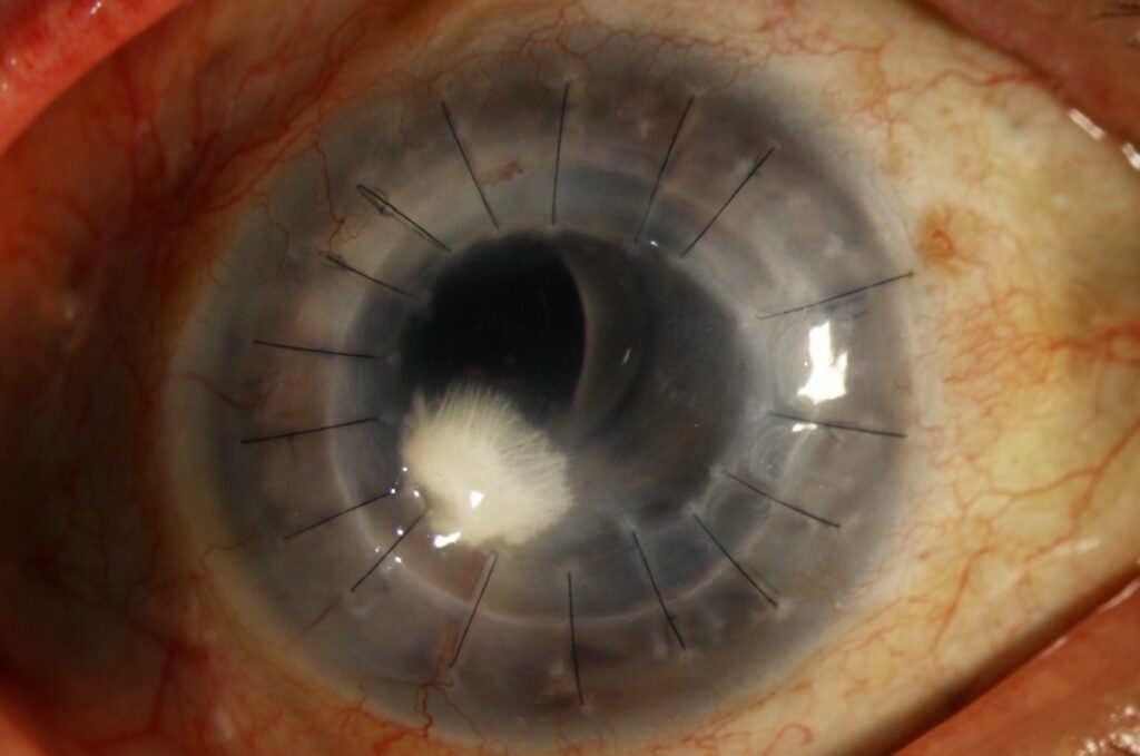
Crystalline keratopathy describes a branching, needle-like pattern of white to gray opacities within the corneal stroma, most often caused by an indolent bacterial infection that grows in a biofilm largely shielded from the host immune response and from topical antibiotics.
It develops almost exclusively in corneas that have had a graft or long-term topical corticosteroid use, and it can smolder for months before it is recognized.
What Causes Crystalline Keratopathy?
The classic organism is Streptococcus viridans, though other streptococci, staphylococci, and occasionally fungi have been implicated.
The bacteria form a biofilm within the corneal stroma, growing along tissue planes in a branching pattern that gives the condition its name, and the biofilm protects the organisms from both antibiotic penetration and the host inflammatory response, which is why the eye often looks relatively quiet despite an active infection.
Chronic topical corticosteroid use, common after corneal transplantation, suppresses the inflammatory response further and allows the infection to progress with minimal symptoms.
Risk Factors
- Penetrating or lamellar corneal transplantation, by far the most common setting
- Long-term topical corticosteroid use
- Chronic bandage contact lens wear
- Previous herpetic keratitis or other causes of corneal epithelial compromise
- Suture-related infection at a loose or exposed suture
All-fit Slit-Lamp Adapter
Record and share exactly what you see at the slit lamp. One adapter fits any slit lamp or surgical microscope — and any smartphone.
From Choroida — the team behind this siteClinical Features
Patients often have few symptoms, since the biofilm shields the process from a brisk inflammatory response.
- Branching, needle-like, white to gray-white opacities in the stroma, often at the graft-host junction
- Minimal surrounding inflammation relative to the depth and extent of the lesion
- Slow progression over weeks to months if untreated
- Reduced vision if the crystalline deposits encroach on the visual axis
The mismatch between the quiet-looking eye and the depth of the infiltrate is itself a clue, since a truly sterile stromal opacity of this pattern is uncommon.
Diagnosis
Corneal scraping and culture from the affected area, ideally obtained before or immediately upon reducing corticosteroid therapy, identifies the organism in many cases, though the biofilm nature of the infection can make culture yield inconsistent.
Confocal microscopy can show the branching bacterial colonies in vivo and support the diagnosis when culture is negative.
A history of graft surgery or chronic steroid use in a patient with a slowly progressive branching stromal opacity should raise this diagnosis even before culture results return.
Management
Reducing Immunosuppression
Topical corticosteroids are tapered or temporarily stopped to allow the host immune response to help clear the infection, balanced against the risk of graft rejection in transplanted corneas.
Antibiotic Therapy
Intensive fortified topical antibiotics are used, chosen to cover streptococcal and staphylococcal organisms, and treatment is often prolonged because the biofilm limits drug penetration.
Some cases require months of therapy, and response is judged by regression of the crystalline pattern rather than by resolution of symptoms, since symptoms were often minimal to begin with.
Surgical Options
Deep lesions or those unresponsive to medical therapy may need therapeutic keratoplasty to remove the infected tissue, particularly when vision is threatened or the infection does not respond to prolonged antibiotic treatment.
Prognosis
Response to treatment can be slow, and recurrence is possible if corticosteroids are resumed too quickly.
Visual outcome depends on the depth and location of the crystalline deposits and whether repeat keratoplasty is needed, but many cases are controlled with medical therapy alone when recognized early.


Document what you see
Two smartphone imaging tools built for everyday clinic use — one for the slit lamp, one for the fundus.
From Choroida — the team behind this siteReferences
- Meisler DM, Langston RH, Naab TJ, Aaby AA, McMahon JT, Tubbs RR. Infectious crystalline keratopathy. Am J Ophthalmol. 1984;97:337-343.
- Fulcher SF, Fader RC, Rosa RH Jr, Holmes GP. Delayed-onset mycobacterial keratitis after LASIK. Cornea. 2002;21:546-554.
- Porter AJ, Lee GA, Jun AS. Infectious crystalline keratopathy. Surv Ophthalmol. 2018;63:480-499.
- Reiss GR, Campbell RJ, Bourne WM. Infectious crystalline keratopathy. Surv Ophthalmol. 1986;31:69-72.
Test yourself
A few questions straight from this article.
-
What corneal appearance defines crystalline keratopathy?
Crystalline keratopathy is a branching, needle-like pattern of white to gray opacities within the corneal stroma, the pattern that gives the condition its name. -
Which organism is the classic cause of infectious crystalline keratopathy?
Streptococcus viridans is the classic organism, although other streptococci, staphylococci and occasionally fungi have also been implicated. -
Why does the eye often look quiet in infectious crystalline keratopathy?
The bacteria grow as a biofilm within the stroma, which protects them from both antibiotic penetration and the host inflammatory response, so the eye looks relatively quiet despite active infection. -
Which setting is by far the most common for infectious crystalline keratopathy?
Penetrating or lamellar corneal transplantation is listed as by far the most common setting, together with long-term topical corticosteroid use. -
Where are the crystalline stromal opacities most often located?
The branching needle-like white to gray-white stromal opacities are often found at the graft-host junction in a transplanted cornea. -
Which test can show branching bacterial colonies in vivo when cultures are negative in crystalline keratopathy?
Confocal microscopy can demonstrate the branching bacterial colonies in vivo and supports the diagnosis when culture yield is inconsistent or negative. -
How are topical corticosteroids handled when treating infectious crystalline keratopathy in a graft?
Steroids are tapered or temporarily stopped so the host immune response can help clear the infection, balanced against the risk of graft rejection in a transplanted cornea. -
How is response to antibiotic therapy judged in infectious crystalline keratopathy?
Response is gauged by regression of the crystalline pattern rather than symptom resolution, because symptoms were usually minimal from the outset. -
Which surgical option is used for deep or medically unresponsive crystalline keratopathy?
Deep lesions, or those not responding to prolonged antibiotics, may require therapeutic keratoplasty to excise the infected tissue, especially when vision is threatened. -
What most commonly precipitates recurrence after treated crystalline keratopathy?
Response to treatment is slow and recurrence is possible if corticosteroids are restarted too quickly after the infection appears controlled.

Writes and reviews the clinical articles on Educate. Every post is checked against peer-reviewed sources before it goes live.


