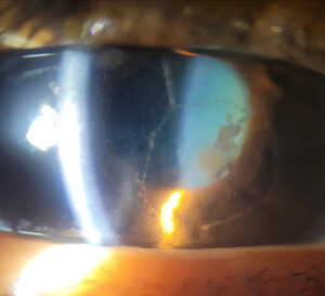
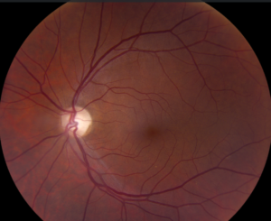
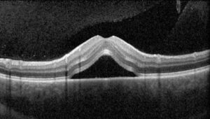
A mulberry lesion is a calcified, multinodular retinal astrocytic tumor with a glistening, lumpy surface.
In addition, most examples are benign hamartomas, and many occur in tuberous sclerosis complex (TSC).
However, retinoblastoma and optic disc drusen can look similar, so careful examination and imaging matter.
What a Mulberry Lesion Is
The tumor arises from astrocytes in the retinal nerve fiber layer.
For example, Nyboer and colleagues described three fundus patterns in TSC: flat translucent, calcified mulberry, and transitional.
Because the clustered nodules resemble the fruit, the calcified form carries its name.
Who Develops a Mulberry Lesion
TSC is the classic association, and it results from TSC1 or TSC2 pathogenic variants.
In addition, retinal hamartomas occur in many patients with TSC, often bilaterally.
However, isolated tumors also occur in otherwise healthy people.
Therefore, a single lesion raises the question of TSC without confirming it.
Fundus Explorer Pro
Photograph the retinal findings described here with the phone already in your pocket — 22 D optics and built-in illumination in one handheld unit.
From Choroida — the team behind this siteSymptoms in Mulberry Lesion
Many patients have no visual symptoms.
Therefore, the finding often appears during screening.
However, vision falls if the tumor involves the fovea or causes a complication.
By contrast, new blur or floaters should prompt a search for exudation, vitreous hemorrhage, or growth.
Fundus Signs of a Mulberry Lesion
Examiners typically find an opaque, yellow-white, nodular mass near the disc or in the posterior pole.
Also, the surface looks refractile because calcified nodules reflect light.
- Well-defined, multilobulated borders
- Location on the inner retinal surface
- Fine intrinsic vessels without large feeding vessels
- Little or no exudation or subretinal fluid
OCT in Mulberry Lesion
First, OCT localizes the tumor to the inner retina and shows its relationship to the fovea.
In addition, Shields and colleagues described a “moth-eaten” pattern of optically empty spaces in many astrocytic hamartomas.
Also, dense calcification casts posterior shadowing, which can hide deeper layers.
Therefore, ultrasound better documents calcium in a thick mass.
Imaging Beyond OCT
Also, calcified nodules often appear brightly hyperautofluorescent, which gives a quick, noninvasive clue.
Next, B-scan ultrasound shows a highly reflective surface with acoustic shadowing.
Mulberry Lesion vs Retinoblastoma
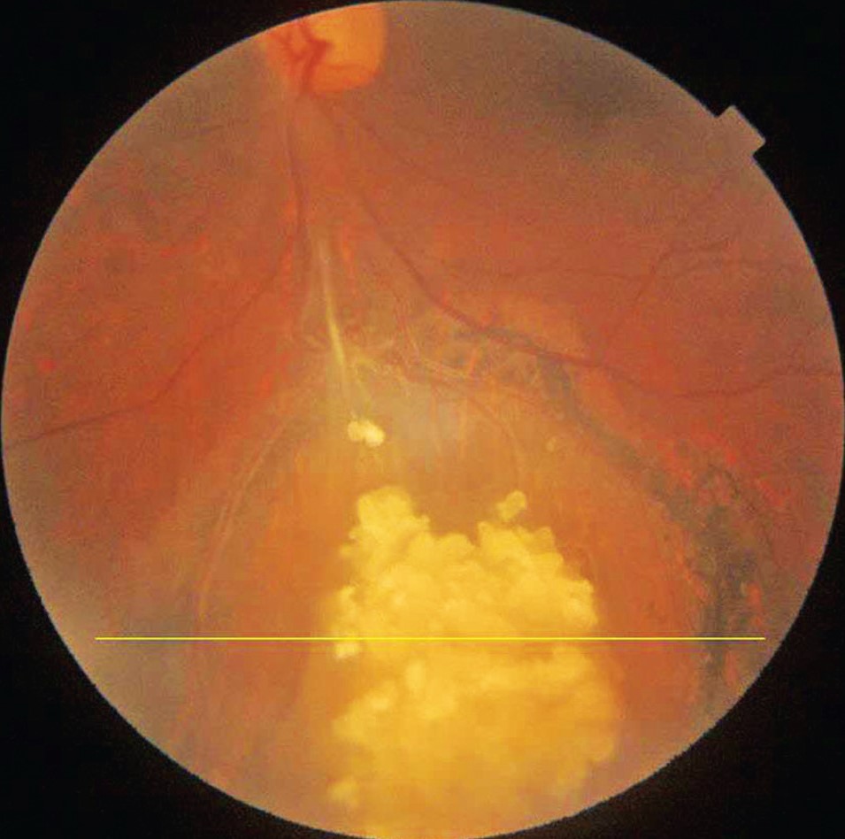
Because retinoblastoma frequently calcifies, calcium alone cannot separate these conditions.
Retinoblastoma typically presents in early childhood with leukocoria or strabismus.
Also, it tends to grow, develop dilated feeding vessels, and shed vitreous or subretinal seeds.
In contrast, a mulberry lesion usually lacks seeding and remains stable.
Therefore, any white intraocular mass in a young child needs urgent ocular oncology assessment.
Mulberry Lesion vs Optic Disc Drusen
First, location gives a clue, because optic disc drusen lie within the optic nerve head.
In addition, they elevate the disc and blur its margins, sometimes causing pseudopapilledema.
In contrast, a mulberry lesion sits on the retinal surface as a discrete mass.
Next, enhanced depth imaging OCT shows drusen as signal-poor bodies with hyperreflective margins.
However, when the disc looks swollen, use a broader approach to optic disc swelling.
Other Mimics
Several conditions can resemble a white or yellow fundus mass:
- For example, myelinated nerve fibers, which show feathery edges
- Also, ocular toxocariasis, which may cause traction
- In addition, Coats disease, which features telangiectasia and exudation
- Finally, choroidal osteoma, which lies deeper
Systemic Workup After a Mulberry Lesion
First, ask about seizures, developmental concerns, renal disease, and family history.
Then inspect the skin for hypomelanotic macules, facial angiofibromas, and ungual fibromas.
Also, current TSC criteria count multiple retinal hamartomas as a major feature.
Consequently, suspected cases need genetic testing and multisystem evaluation.
Follow-Up Plan
First, baseline documentation should include color photography, OCT, and autofluorescence when available.
Next, guidelines recommend a baseline dilated examination in TSC, with repeat assessment according to findings.
However, review earlier when the tumor sits near the fovea or optic nerve.
Finally, compare size, surface, fluid, and vitreous findings with previous images at each visit.
Treatment of a Complicated Mulberry Lesion
Because treatment offers no clear benefit, observation suits most stable, asymptomatic lesions.
However, progressive exudation, growth, or neovascular complications may justify intervention.
Reported options include laser, photodynamic therapy, anti-VEGF injections, and systemic mTOR inhibitors.
As a result, benefit remains uncertain, because evidence comes mainly from small series and case reports.
In addition, these treatments cannot reverse established foveal damage.
Counseling the Patient
First, explain that the tumor is usually benign and often needs monitoring alone.
Finally, patients should report new blur, floaters, or field changes promptly.
Pitfalls to Avoid
- Do not treat calcification as proof of a benign lesion
- Never diagnose a mulberry lesion in a child without excluding retinoblastoma
- Also avoid confusing a surface tumor with buried disc drusen


Document what you see
Two smartphone imaging tools built for everyday clinic use — one for the slit lamp, one for the fundus.
From Choroida — the team behind this siteReferences
- Nyboer JH, Robertson DM, Gomez MR. Retinal lesions in tuberous sclerosis. Arch Ophthalmol. 1976;94:1277-1280.
- Aronow ME, Nakagawa JA, Gupta A, Traboulsi EI, Singh AD. Tuberous sclerosis complex: genotype/phenotype correlation of retinal findings. Ophthalmology. 2012;119:1917-1923.
- Shields CL, Say EAT, Fuller T, Arora S, Samara WA, Shields JA. Retinal astrocytic hamartoma arises in nerve fiber layer and shows “moth-eaten” optical coherence tomographic pattern. Ophthalmology. 2016;123:1809-1816.
- Northrup H, Aronow ME, Bebin EM, et al. Updated international tuberous sclerosis complex diagnostic criteria and surveillance and management recommendations. Pediatr Neurol. 2021;123:50-66.
- Palmer E, Gale J, Crowston JG, Wells AP. Optic nerve head drusen: an update. Neuroophthalmology. 2018;42:367-384.

Writes and reviews the clinical articles on Educate. Every post is checked against peer-reviewed sources before it goes live.