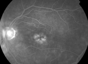
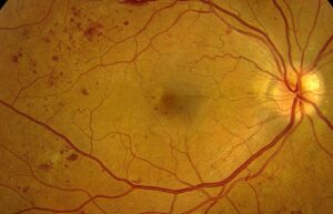
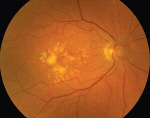
Keith-Wagener-Barker classification sorts hypertensive fundus findings into four groups of rising severity.
Keith, Wagener and Barker published it in 1939 after following patients with hypertension.
It still appears in textbooks, although simpler schemes now compete with it.
Why Keith-Wagener-Barker Was Developed
The authors wanted to connect fundus appearance with the clinical course of hypertension.
Therefore, they grouped patients by ophthalmoscopic findings and followed survival.
Effective antihypertensive drugs were not yet available, so the data reflect largely untreated disease.
The Four Keith-Wagener-Barker Groups
Each group builds on the findings of the one before it.
- Mild generalized arteriolar narrowing defines group I
- More marked narrowing, focal constriction and arteriovenous crossing changes define group II
- Next, group III adds hemorrhages, cotton wool spots and hard exudates
- Finally, optic disc swelling defines group IV
Fundus Explorer Pro
Photograph the retinal findings described here with the phone already in your pocket — 22 D optics and built-in illumination in one handheld unit.
From Choroida — the team behind this siteVessel Changes in Groups I and II
Sustained pressure first causes arteriolar vasoconstriction.
Later, intimal thickening and medial hyperplasia make the vessel wall more opaque.
The examiner may then see copper wiring and venous compression at crossings.
Most patients report no visual symptoms at this stage.
Barrier Breakdown in Groups III and IV
Higher grades reflect damage to the blood-retinal barrier and nerve fiber layer ischemia.
Flame hemorrhages follow the nerve fiber layer, while cotton wool spots mark focal axoplasmic stasis.
Hard exudates may also form a macular star, and vision can then fall.
Disc swelling should prompt urgent assessment for hypertensive emergency.
Examining Keith-Wagener-Barker Signs
A dilated examination with a fundus lens gives the best view of the posterior pole.
First, compare arteriolar and venular caliber away from the disc.
Then, inspect crossings for nicking and scan the nerve fiber layer for hemorrhages.
OCT can confirm macular fluid or disc edema when the picture looks uncertain.
Limits of Keith-Wagener-Barker Grading
Groups I and II are hard to separate, because narrowing varies and judgment remains subjective.
In addition, age and arteriosclerosis can produce similar vessel changes.
The sequence also implies steady progression, although severe pressure can cause exudative signs without long-standing vessel disease.
Therefore, a fundus grade cannot show how long hypertension has been present.
Older and Newer Grading Schemes
Scheie graded hypertensive changes and arteriolar sclerosis separately in 1953.
Wong and Mitchell later proposed a simpler scheme of none, mild, moderate and malignant retinopathy.
Mild disease includes arteriolar narrowing, nicking and wall opacification.
Moderate disease adds hemorrhages, microaneurysms, cotton wool spots or hard exudates.
Malignant disease then adds optic disc swelling.
Dodson and colleagues also suggested simplified grading because early vessel grades separate poorly.
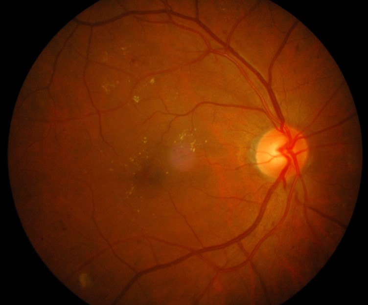
Keith-Wagener-Barker Versus Modern Schemes
Modern schemes essentially merge groups I and II into one mild category.
Groups III and IV map closely to the moderate and malignant categories.
Either system works if the note also describes the actual signs.
Prognosis Beyond Keith-Wagener-Barker
Survival fell steeply from group I to group IV in the 1939 report, but modern treatment has changed this course.
Population studies now offer better prognostic data.
For example, the ARIC study linked retinal microvascular abnormalities with incident stroke after accounting for other risk factors.
However, these figures describe groups, so they cannot predict one patient’s outcome.
What Keith-Wagener-Barker Leaves Out
The system focuses on retinal vessels and the disc, so it does not grade the choroid.
Yet acute hypertension can also cause hypertensive choroidopathy, with Elschnig spots and serous detachments.
Later, Siegrist streaks may mark earlier choroidal damage.
What Must Be Excluded
Hemorrhages and cotton wool spots are nonspecific signs.
Therefore, consider diabetic retinopathy, retinal vein occlusion, anemia, HIV retinopathy and radiation damage.
Bilateral, fairly symmetric findings with high blood pressure favor hypertension.
Similarly, disc swelling requires you to consider raised intracranial pressure and optic neuritis.
Management by Keith-Wagener-Barker Grade
The grade guides urgency, while the physician treats the underlying pressure.
Patients with vessel changes alone usually need a blood pressure check and primary care follow-up.
In contrast, severe retinopathy with disc swelling requires same-day medical evaluation.
Controlled lowering matters, because abrupt reduction can cause optic nerve and brain ischemia.
Retinal signs often regress with treatment, though disc pallor or macular damage may limit recovery.
Explain to the patient that the retina offers a direct view of small vessels.
See hypertensive retinopathy for a broader review.
Pitfalls to Avoid
- Do not quote 1939 survival data as a current prognosis
- Avoid attributing every hemorrhage to hypertension without checking glucose and blood count
- Never send a patient with disc swelling home without a blood pressure reading
- Do not record a group number without describing the signs


Document what you see
Two smartphone imaging tools built for everyday clinic use — one for the slit lamp, one for the fundus.
From Choroida — the team behind this siteReferences
- Keith NM, Wagener HP, Barker NW. Some different types of essential hypertension: their course and prognosis. Am J Med Sci. 1939;197:332-343.
- Scheie HG. Evaluation of ophthalmoscopic changes of hypertension and arteriolar sclerosis. AMA Arch Ophthalmol. 1953;49:117-138.
- Wong TY, Mitchell P. Hypertensive retinopathy. N Engl J Med. 2004;351:2310-2317.
- Dodson PM, Lip GY, Eames SM, Gibson JM, Beevers DG. Hypertensive retinopathy: a review of existing classification systems and a suggestion for a simplified grading system. J Hum Hypertens. 1996;10:93-98.
- Wong TY, Klein R, Couper DJ, et al. Retinal microvascular abnormalities and incident stroke: the Atherosclerosis Risk in Communities Study. Lancet. 2001;358:1213-1217.

Writes and reviews the clinical articles on Educate. Every post is checked against peer-reviewed sources before it goes live.