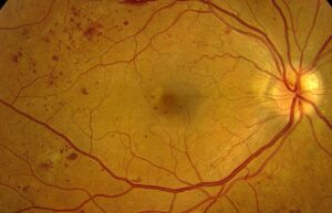
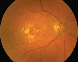
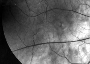
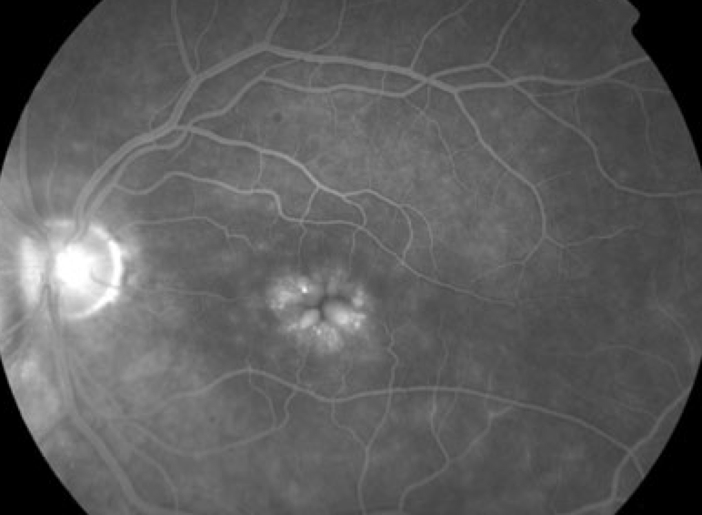
Petaloid leakage is the flower-shaped late hyperfluorescence that fluorescein angiography (FA) shows in classic cystoid macular edema (CME).
Dye collects in cystoid spaces around the fovea and outlines each petal.
However, several cystic maculopathies never leak, so the sign needs careful interpretation.
What Petaloid Leakage Means
The sign reflects dye pooling after breakdown of the inner blood-retinal barrier.
Perifoveal capillaries usually supply the leak.
Fluorescein then escapes into the retina and fills cystoid spaces.
As a result, the examiner sees a rosette of bright petals around the fovea.
The Anatomy Behind the Flower
Henle fibers leave the foveal cones and run obliquely outward in a radial array.
Müller cell processes also form partitions in this region.
Therefore, fluid in the outer plexiform layer collects in elongated compartments that point toward the foveal center.
Each compartment then fills with dye and forms one petal.
Fundus Explorer Pro
Photograph the retinal findings described here with the phone already in your pocket — 22 D optics and built-in illumination in one handheld unit.
From Choroida — the team behind this sitePetaloid Leakage Versus a Honeycomb Pattern
Farther from the fovea, the fibers lose this radial arrangement.
Cysts in the inner nuclear layer then tend to form a polygonal honeycomb pattern.
In addition, large central cysts can merge and blur the individual petals.
Timing of Petaloid Leakage on FA
Early frames may look nearly normal or show tiny perifoveal points of hyperfluorescence.
Midphase frames then show dye spreading into the retina.
Finally, the full flower usually matures in the late frames, several minutes after injection.
Reading the Late Frames
True leakage grows in area and intensity while its borders blur.
Staining keeps its borders, whereas a window defect fades with the background (see leakage, blockage and staining).
Also inspect the disc, because disc leakage may accompany pseudophakic or inflammatory CME.
Common Causes of Petaloid Leakage
Pseudophakic CME (Irvine-Gass syndrome) gives the textbook picture.
Uveitis, retinal vein occlusion and diabetic macular edema can also produce it.
Drug associations also exist, for example fingolimod and, with less certainty, topical prostaglandin analogs after cataract surgery.
Clues to the Underlying Cause
The rest of the angiogram often identifies the source.
For example, microaneurysms and capillary dropout favor diabetic disease.
Sectoral hemorrhage, collaterals and nonperfusion suggest vein occlusion.
Meanwhile, fern-like peripheral vessel leakage points toward uveitis (see leaky vessels on angiography).
Petaloid Leakage on Exam and OCT
OCT shows hyporeflective cysts, often with subretinal fluid in the central macula.
It measures thickness well, yet it cannot directly show barrier leakage.
Similarly, OCT angiography maps capillary changes but does not image dye leakage.
Cystic Maculopathy Without Petaloid Leakage
Clear OCT cysts with a silent angiogram should change your reasoning.
In these eyes, fluid may come from Müller cell dysfunction, traction or tissue loss.
Barrier-directed treatment therefore has a weaker rationale.
Inherited, Tractional and Drug Causes
X-linked retinoschisis causes foveal schisis, typically without leakage in a young male.
Enhanced S-cone syndrome can also show cystic change with a quiet angiogram.
In addition, vitreomacular traction and epiretinal membrane may create tractional cysts.
Nicotinic acid and taxanes can also cause cysts with little leakage.
Microcystic change from optic neuropathy and foveal cavitation in macular telangiectasia type 2 can also mimic edema.
When to Order FA
OCT usually confirms macular edema, so not every patient needs dye.
FA helps when the cause is unclear or the response to therapy is poor.
It also helps when you suspect vasculitis, ischemia or a silent cystic maculopathy.
Before injection, warn patients about nausea and yellow skin and urine, and mention rare serious allergic reactions.
How Petaloid Leakage Changes Management
A positive study supports treatment directed at the cause and the barrier breakdown.
For example, clinicians often use topical NSAIDs and corticosteroids for pseudophakic CME, although trial evidence has limitations.
Uveitic and vascular edema may need corticosteroids or anti-VEGF injections, each with its own risks.
However, none of these treatments can restore photoreceptors already lost to chronic edema.
Follow-Up After Petaloid Leakage
OCT then becomes the main monitoring tool, because it is quick and needs no injection.
Track visual acuity, central thickness and outer retinal integrity at each visit.
Angiographic leakage can also persist after vision improves, so it should not drive treatment alone.
Counseling the Patient
Explain that tiny macular vessels are leaking fluid into the central retina.
Many pseudophakic cases improve, while chronic edema may leave permanent distortion or reduced acuity.
Finally, ask patients to report new blur or distortion early.
Pitfalls to Avoid
- Do not end the study before the late macular frames
- Never assume that every OCT cyst represents leaking CME
- Avoid escalating injections in a silent cystic maculopathy
- Do not overlook niacin or taxane use in the history
- Remember that media opacity can obscure a subtle flower


Document what you see
Two smartphone imaging tools built for everyday clinic use — one for the slit lamp, one for the fundus.
From Choroida — the team behind this siteReferences
- Gass JD, Norton EW. Cystoid macular edema and papilledema following cataract extraction: a fluorescein fundoscopic and angiographic study. Arch Ophthalmol. 1966;76:646-661.
- Yanoff M, Fine BS, Brucker AJ, Eagle RC Jr. Pathology of human cystoid macular edema. Surv Ophthalmol. 1984;28 Suppl:505-511.
- Johnson MW. Etiology and treatment of macular edema. Am J Ophthalmol. 2009;147:11-21.
- Spaide RF. Retinal vascular cystoid macular edema: review and new theory. Retina. 2016;36:1823-1842.
- Gass JD. Nicotinic acid maculopathy. Am J Ophthalmol. 1973;76:500-510.

Writes and reviews the clinical articles on Educate. Every post is checked against peer-reviewed sources before it goes live.