NPDR vs PDR is the main staging question in diabetic retinopathy, and neovascularization separates the two stages.
NPDR describes microvascular damage within the retina, while PDR adds new vessels driven by ischemia.
Therefore, this boundary changes risk, follow-up and treatment.
NPDR vs PDR: The Defining Difference
NPDR has no neovascularization, and its lesions stay within the retina.
In contrast, PDR requires new vessels on the disc or elsewhere, or hemorrhage arising from them.
The ICDR and ETDRS scales both use this boundary (see diabetic retinopathy classification).
Fundus Findings in NPDR
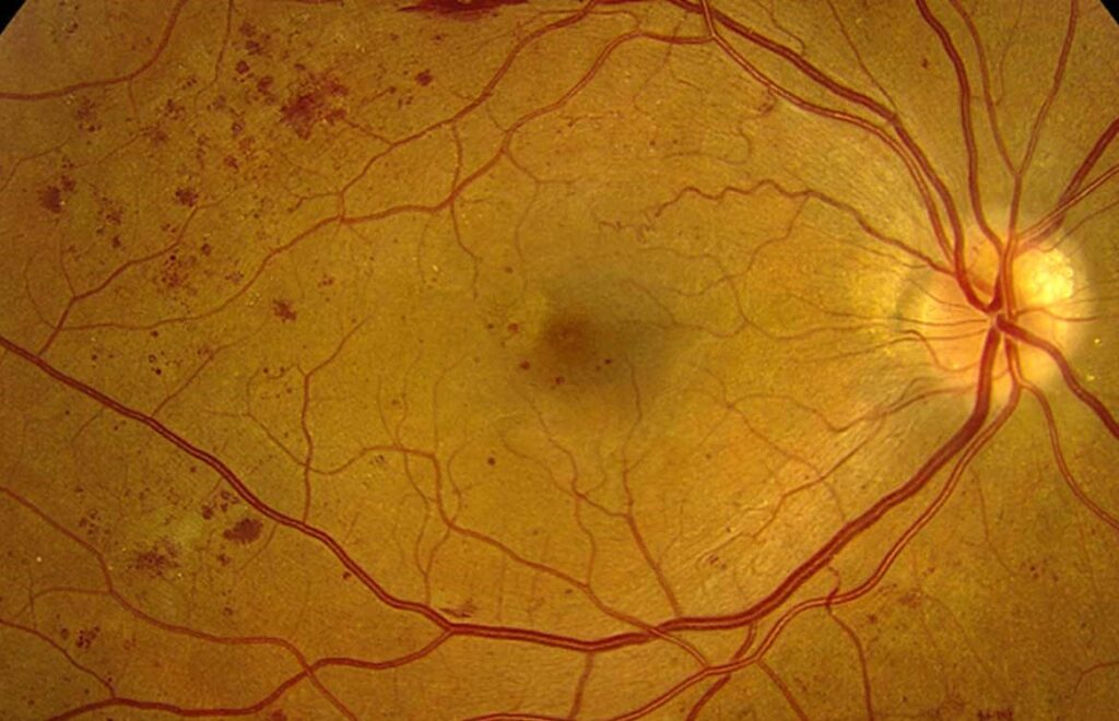
Microaneurysms appear first, and they may be the only sign in mild disease.
Then, dot and blot hemorrhages, hard exudates and cotton wool spots appear as damage advances.
Venous beading and intraretinal microvascular abnormalities (IRMA) suggest significant ischemia.
Fundus Explorer Pro
Photograph the retinal findings described here with the phone already in your pocket — 22 D optics and built-in illumination in one handheld unit.
From Choroida — the team behind this siteFundus Findings in PDR
New vessels form fine, irregular loops on the disc (NVD) or elsewhere (NVE).
They often bleed when the vitreous pulls on them.
Later, fibrovascular tissue may contract and cause tractional retinal detachment.
In addition, check the iris and angle before dilation, because neovascularization there signals severe ischemia.
Severe NPDR and the 4-2-1 Rule
Severe NPDR marks the last stage before neovascularization.
The rule requires one of the following, with no signs of PDR:
- Severe intraretinal hemorrhages in all four quadrants
- Definite venous beading in at least two quadrants
- Prominent IRMA in at least one quadrant
High-Risk PDR
The Diabetic Retinopathy Study defined the features that carry the greatest threat to vision.
High-risk PDR includes NVD of about one quarter to one third disc area or more.
It also includes any NVD with vitreous or preretinal hemorrhage.
Imaging NPDR vs PDR
Fluorescein angiography shows nonperfusion in both stages, while active new vessels leak profusely.
As a result, it helps separate NVE from IRMA, which leaks little.
OCT angiography can show flow above the internal limiting membrane without dye.
However, consider retinal vein occlusion or ocular ischemic syndrome when findings look asymmetric.
Examination Pearls in NPDR vs PDR
First, examine the dilated fundus with a 90 diopter lens and scan all four quadrants.
Next, look for NVE along the arcades, because it often hides near the vessels.
Also, use red-free light to separate fine new vessels from IRMA.
Finally, ask the patient to look in different directions, because the periphery may reveal early traction.
Differential Diagnosis of Neovascularization
Several diseases can mimic diabetic neovascularization, so the history matters.
For example, retinal vein occlusion, sickle cell retinopathy and ocular ischemic syndrome can all produce new vessels.
In addition, a normal glucose history should prompt a search for these causes.
Therefore, confirm the diagnosis before you label an eye as PDR.
Progression Risk in NPDR vs PDR
ETDRS data showed that risk climbs steeply as NPDR severity increases.
For example, severe NPDR carries about a 15 percent one-year risk of high-risk PDR.
Very severe NPDR raises that figure to about 45 percent.
Follow-Up Intervals: NPDR vs PDR
Intervals shorten as severity increases, although guidelines allow individual adjustment.
Mild NPDR usually needs review about yearly, while severe NPDR generally needs review within 3 to 4 months.
Active or high-risk PDR needs prompt treatment.
Treatment Thresholds in NPDR
Systemic control of glucose, blood pressure and lipids forms the basis of NPDR care.
Most eyes without macular edema then need observation only.
PANORAMA showed that aflibercept reduced progression to vision-threatening complications in moderately severe to severe NPDR.
However, Protocol W found no visual acuity advantage at four years with preventive treatment (see anti-VEGF for NPDR).
Treatment Thresholds in PDR
High-risk PDR warrants prompt treatment, because the Diabetic Retinopathy Study showed that PRP cuts severe vision loss by at least 50 percent.
PRP can cause peripheral field loss, reduced night vision and worsened macular edema.
Protocol S found ranibizumab noninferior to PRP for visual acuity at two years.
However, anti-VEGF needs repeated visits (see PRP, anti-VEGF, or both).
Vitrectomy then addresses nonclearing hemorrhage or traction threatening the macula.
Counseling on NPDR vs PDR
Both stages can remain silent, so good acuity never excludes neovascularization.
Explain that PDR means abnormal new vessels that can bleed.
Then, describe the warning symptoms: new floaters, a dark curtain or sudden blur.
Patients choosing anti-VEGF should understand that missed visits can allow rapid recurrence.
Pitfalls to Avoid
- Do not call IRMA neovascularization without checking its location and leakage
- Never skip gonioscopy when ischemia looks severe
- Do not rely on visual acuity to judge stage
- Avoid anti-VEGF alone for patients with unreliable follow-up


Document what you see
Two smartphone imaging tools built for everyday clinic use — one for the slit lamp, one for the fundus.
From Choroida — the team behind this siteReferences
- Flaxel CJ, Adelman RA, Bailey ST, et al. Diabetic Retinopathy Preferred Practice Pattern. Ophthalmology. 2020;127:P66-P145.
- Early Treatment Diabetic Retinopathy Study Research Group. Fundus photographic risk factors for progression of diabetic retinopathy: ETDRS report number 12. Ophthalmology. 1991;98:823-833.
- Diabetic Retinopathy Study Research Group. Photocoagulation treatment of proliferative diabetic retinopathy: DRS report number 8. Ophthalmology. 1981;88:583-600.
- Gross JG, Glassman AR, Jampol LM, et al. Panretinal photocoagulation vs intravitreous ranibizumab for proliferative diabetic retinopathy. JAMA. 2015;314:2137-2146.
- Brown DM, Wykoff CC, Boyer D, et al. Evaluation of intravitreal aflibercept for the treatment of severe nonproliferative diabetic retinopathy: the PANORAMA randomized clinical trial. JAMA Ophthalmol. 2021;139:946-955.
- Maturi RK, Glassman AR, Josic K, et al. Four-year visual outcomes in the Protocol W randomized trial of intravitreous aflibercept for prevention of vision-threatening complications of diabetic retinopathy. JAMA. 2023;329:376-385.

Writes and reviews the clinical articles on Educate. Every post is checked against peer-reviewed sources before it goes live.