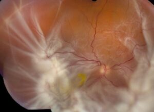
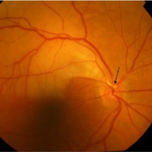
Meridional folds are small radial ridges of retina that lie in line with the dentate processes at the ora serrata.
They are a normal variant, and many clinicians first see them during scleral depression in patients with floaters or flashes.
Knowing their appearance helps to avoid unnecessary worry and treatment.
What Meridional Folds Look Like
The folds appear as short radial elevations of the peripheral retina, often superonasal, that point toward the ora serrata.
They may be single or multiple, and they often end in a small white notch or a pigment spot at the anterior end.
Spencer, Foos, and Straatsma described these structures and their associations in a classic autopsy study.
On indirect ophthalmoscopy they look like folded retinal tissue lying in the line of a dentate process (see peripheral retinal variants mimicking pathology).
How Common Meridional Folds Are
Studies report folds in a minority of eyes, and they are more common in the superior and nasal quadrants.
They are present from birth, and they do not change over time in most eyes.
They are usually bilateral, and they are not associated with symptoms.
Fundus Explorer Pro
Photograph the retinal findings described here with the phone already in your pocket — 22 D optics and built-in illumination in one handheld unit.
From Choroida — the team behind this siteFindings Related to Meridional Folds
Meridional complexes are folds that connect with a dentate process or an enclosed bay.
Spencer and colleagues found that some folds had a retinal tear at the posterior end, though the risk is low.
Look for retinal breaks around the fold edge, especially when symptoms such as flashes or new floaters appear (see horseshoe tears).
Differential Diagnosis of Meridional Folds
- Retinal tears and giant tears at the ora
- Lattice degeneration and retinal tufts
- Retinal dialysis, which is a break at the ora
- Retinoschisis and peripheral cysts
Careful scleral depression and good illumination separate these (see lattice degeneration).
Management of Meridional Folds
Do not treat meridional folds with laser or cryotherapy, since the folds themselves do not threaten the retina.
Treat only associated retinal tears that show traction or symptoms.
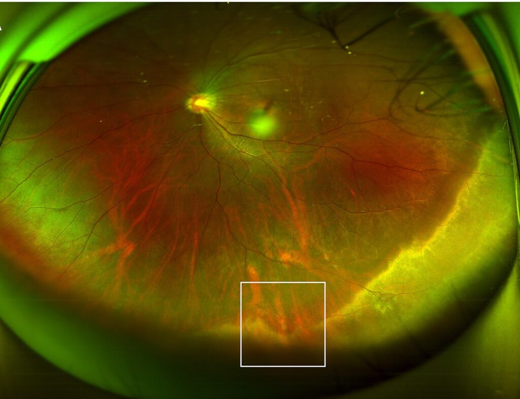
Document the finding with a drawing or wide-field photograph, so that future examiners recognize it.
Educate patients about symptoms of detachment, as for any patient with floaters.
Counselling About Meridional Folds
Explain that the folds are a normal variation and not a sign of disease.
Reassure patients that the risk of retinal detachment from the folds themselves is very low.
Advise them to return for new flashes, new floaters, or a shadow in the vision.
An Example Case
A 31-year-old man reports a few new floaters in his right eye, and the dilated examination is otherwise normal.
Scleral depression shows a short radial ridge in the superonasal retina, ending at a small white notch next to the ora serrata.
The ridge is stable, and no tear is present around it.
The doctor documents the fold with a drawing and reassures the patient.
Follow-up a year later shows no change.
Questions Patients Ask
Patients ask whether the fold will cause a detachment, and the risk from the fold alone is very low.
They also ask whether they need laser, and the answer is no.
Because the finding looks unusual, show the patient the drawing and explain that it is a normal variant.
Pitfalls to Avoid
- Treating a normal fold with laser or cryotherapy
- Missing a retinal tear at the posterior end of the fold
- Calling every peripheral ridge a fold without checking its position
- Forgetting to record the finding for future visits
Looking Ahead
Wide-field imaging may help to document folds and their variants.
Also, OCT of the periphery may show their structure more clearly.
Therefore, future clinicians may separate them from pathology with less uncertainty.
Practical Points
Use scleral depression with careful illumination on every dilated peripheral exam.
Then compare the fold with the dentate process, since they share a line.
Also document the location in clock hours.
Related Normal Variants
Other benign findings include pars plana cysts, enclosed oral bays, and peripheral cystoid degeneration.
Learning these variants reduces the risk of overtreatment.
Therefore, review the normal anatomy of the ora serrata regularly.
Why the Distinction Matters
Many patients seek care because of floaters or flashes, and a benign fold may draw attention away from a true tear.
Therefore, examine the whole periphery for 360 degrees.
Also use scleral depression, because folds and tears can look alike without it.
Then record each finding separately.
Follow-Up
Review the patient after a few weeks if symptoms persist.
Because a posterior vitreous detachment can create new breaks, repeat the examination when symptoms change.
Teaching Residents
Show residents how a fold lines up with a dentate process.
Then ask them to draw the fold and to name the nearby landmarks.
Because practice builds confidence, repeat the exercise during each rotation.
For the latest studies, see the PubMed results for meridional folds.


Document what you see
Two smartphone imaging tools built for everyday clinic use — one for the slit lamp, one for the fundus.
From Choroida — the team behind this siteReferences
- Spencer LM, Foos RY, Straatsma BR. Meridional folds, meridional complexes, and associated abnormalities of the peripheral retina. Am J Ophthalmol. 1970;70:697-714.
- Byer NE. The natural history of asymptomatic retinal breaks. Ophthalmology. 1982;89:1033-1039.
- Lewis H. Peripheral retinal degenerations and the risk of retinal detachment. Am J Ophthalmol. 2003;136:155-160.
- American Academy of Ophthalmology. Basic and Clinical Science Course, Section 12: Retina and Vitreous. AAO; current edition.

Writes and reviews the clinical articles on Educate. Every post is checked against peer-reviewed sources before it goes live.