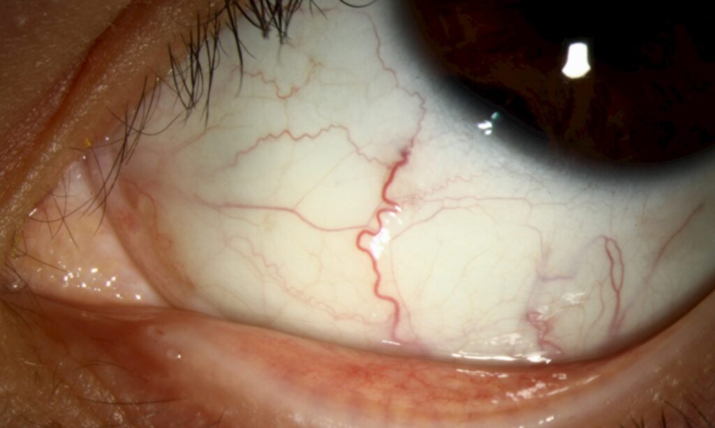
Sentinel vessels are dilated, tortuous episcleral vessels that cluster over one quadrant of the globe.
In an adult, they often overlie a ciliary body melanoma hidden behind the iris. Therefore, a single engorged vessel deserves a dilated examination of the same quadrant.
What Sentinel Vessels Look Like
They appear as one to several thick, dark red vessels on the episclera, and most run toward the limbus and point to a mass hidden behind the iris (see ciliary body melanoma). They also tend to persist after topical phenylephrine, unlike superficial conjunctival injection.
Why Sentinel Vessels Form
A growing ciliary body tumor demands a large blood supply, and as a result, anterior ciliary vessels over the mass dilate to carry the extra flow. However, the sign usually points to the quadrant where the tumor sits.
All-fit Slit-Lamp Adapter
Record and share exactly what you see at the slit lamp. One adapter fits any slit lamp or surgical microscope — and any smartphone.
From Choroida — the team behind this sitePatients Who Need a Closer Look
Ciliary body melanoma mostly affects adults in middle and older age, and a sectoral cataract or new astigmatism in one eye needs a closer look. Children rarely develop melanoma, so a ciliary body mass in a child suggests other tumors (see medulloepithelioma).
Examining the Quadrant
First, dilate the pupil widely, then look behind the iris in the quadrant under the vessels. A brown or amelanotic dome may appear at the pupil margin or in the far periphery. Gonioscopy also checks for tumor in the angle and for iris root displacement.
Transillumination for Sentinel Vessels
Transillumination gives quick, cheap information at the bedside, so darken the room and apply a bright light directly to the sclera, or shine it through the pupil. A pigmented tumor casts a sharp shadow over its base, while amelanotic tumors transmit light and may look falsely reassuring.
UBM in Sentinel Vessels Workup
Ultrasound biomicroscopy (UBM) images the ciliary body at high resolution. A melanoma shows a solid mass with medium to low internal reflectivity, along with angle invasion, ciliochoroidal spread and contact with the lens. A cyst, in contrast, appears hollow with thin walls (see ciliary body cyst).
B-Scan and Other Imaging
B-scan ultrasound measures thickness and posterior extension into the choroid. Anterior segment OCT shows the angle well but penetrates pigmented tissue poorly, and therefore it adds little for thick ciliary body tumors.
Differential Diagnosis of Sentinel Vessels
- Episcleritis: sectoral redness, mild discomfort, often recurrent and self-limited
- Scleritis: deep pain, violaceous hue, tenderness on touch
- Carotid cavernous fistula: corkscrew vessels in all quadrants, often with chemosis
- Previous surgery or trauma: dilated vessels at an old wound
- Other ciliary body tumors: metastasis, adenoma, medulloepithelioma
What Changes the Diagnosis
Pain, diffuse injection and response to steroids favor inflammation. However, a partial response does not exclude a tumor, so image the quadrant anyway. A normal dilated exam with normal UBM makes melanoma unlikely, so photograph the vessels and recheck the quadrant at a later visit.
Steps After Detecting Sentinel Vessels
- Dilated exam with gonioscopy of the same quadrant
- Transillumination and photography of the vessels
- UBM first, then B-scan for posterior extent
- Referral to an ocular oncologist for any solid mass
- Systemic staging, including liver imaging, once melanoma is likely
Staging and Prognosis
Ciliary body involvement upstages a uveal melanoma in the AJCC system and also carries a worse prognosis in large clinical series. Next, genetic testing of biopsy tissue, such as gene expression profiling, refines individual risk. Therefore, early recognition of sentinel vessels may allow treatment of a smaller tumor.
Treatment Options
Plaque brachytherapy treats many small and medium tumors while preserving the eye, and proton beam radiation suits some tumors near the angle or in awkward positions. Local resection can remove selected tumors, although it risks hemorrhage and retinal detachment, while large tumors or blind painful eyes usually need enucleation. Radiation cannot restore lost vision and often causes cataract, dry eye or late radiation damage.
Counseling the Patient With Sentinel Vessels
Explain that the red vessel itself is harmless, although it may point to a tumor. Most patients worry about the eye, while the main threat is spread to the liver, so stress lifelong systemic surveillance even after successful local treatment.
Follow-Up of Sentinel Vessels
After treatment, the vessels may shrink as the tumor regresses. However, persistent or new vessels prompt repeat UBM to check for growth.
Pitfalls to Avoid
- Do not label a single dilated vessel as episcleritis without dilating the pupil
- Never skip gonioscopy of the involved quadrant
- Do not trust a negative transillumination test in an amelanotic tumor
- Avoid repeated topical steroids for unexplained sectoral redness
- Do not delay systemic staging once melanoma is confirmed


Document what you see
Two smartphone imaging tools built for everyday clinic use — one for the slit lamp, one for the fundus.
From Choroida — the team behind this siteReferences
- Shields CL, Kaliki S, Furuta M, et al. Clinical spectrum and prognosis of uveal melanoma based on age at presentation in 8,033 cases. Retina. 2012;32:1363-1372.
- Pavlin CJ, Harasiewicz K, Sherar MD, Foster FS. Clinical use of ultrasound biomicroscopy. Ophthalmology. 1991;98:287-295.
- Kaliki S, Shields CL. Uveal melanoma: relatively rare but deadly cancer. Eye (Lond). 2017;31:241-257.
- Collaborative Ocular Melanoma Study Group. The COMS randomized trial of iodine 125 brachytherapy for choroidal melanoma: V. Twelve-year mortality rates and prognostic factors. Arch Ophthalmol. 2006;124:1684-1693.
- Salmon JF. Kanski’s Clinical Ophthalmology: A Systematic Approach. 9th ed. Elsevier; 2020.
Test yourself
A few questions straight from this article.
-
In an adult, sentinel episcleral vessels often overlie which hidden lesion?
Sentinel vessels are dilated, tortuous episcleral vessels over one quadrant that often overlie a ciliary body melanoma hidden behind the iris. -
Why do sentinel vessels form over a ciliary body tumor?
A growing ciliary body tumor demands a large blood supply, so anterior ciliary vessels over the mass dilate, pointing to the tumor's quadrant. -
What is the key step when a single engorged sectoral episcleral vessel is found in an adult?
Dilate widely and look behind the iris in the quadrant under the vessels, with gonioscopy for angle tumor; never label it episcleritis without dilating. -
A ciliary body mass under sentinel vessels in a child suggests which kind of tumor?
Ciliary body melanoma mostly affects middle-aged and older adults; children rarely develop it, so a mass in a child suggests tumors such as medulloepithelioma. -
Why can transillumination be falsely reassuring when sentinel vessels are present?
A pigmented tumor casts a sharp shadow over its base, while amelanotic tumors transmit light and may look falsely reassuring. -
On ultrasound biomicroscopy, how does a ciliary body melanoma differ from a ciliary body cyst?
A melanoma shows a solid mass with medium to low internal reflectivity, sometimes with angle invasion or lens contact, whereas a cyst appears hollow with thin walls. -
Which treatment preserves the eye for many small and medium ciliary body melanomas?
Plaque brachytherapy treats many small and medium tumors while preserving the eye; large tumors or blind painful eyes usually need enucleation. -
Which feature points to a carotid cavernous fistula rather than sentinel vessels?
A carotid cavernous fistula gives corkscrew vessels in all quadrants, often with chemosis, unlike the sectoral cluster of sentinel vessels. -
How does ciliary body involvement affect a uveal melanoma?
Ciliary body involvement upstages uveal melanoma and carries a worse prognosis, so early recognition of sentinel vessels may allow treatment of a smaller tumor. -
After treatment of a ciliary body melanoma found via sentinel vessels, what is the main lifelong threat to stress?
Most patients worry about the eye, but the main threat is liver spread, so lifelong systemic surveillance continues even after successful local treatment.

Writes and reviews the clinical articles on Educate. Every post is checked against peer-reviewed sources before it goes live.