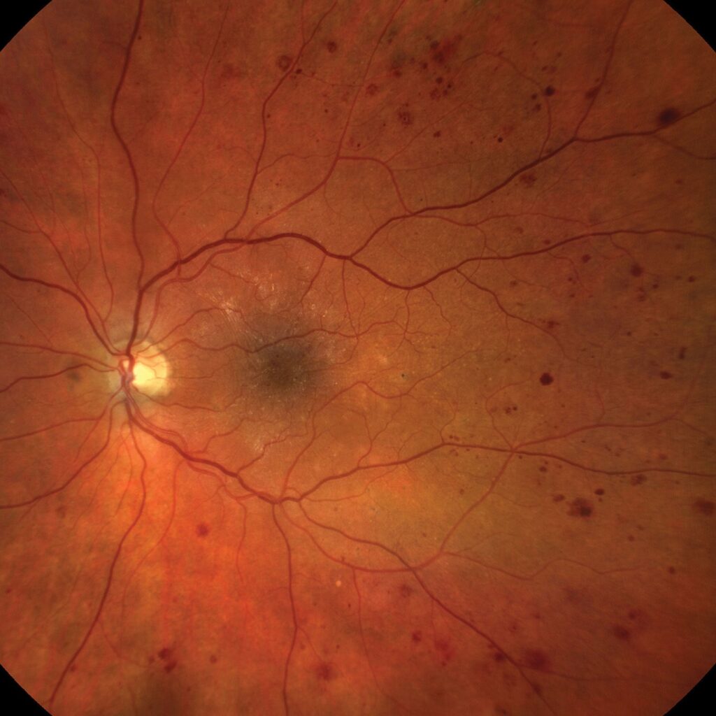
Microaneurysm vs hemorrhage is the first comparison an examiner makes when red dots appear in a diabetic fundus.
Both lesions look small, round and red, but they differ in origin, behavior and grading weight.
What a Microaneurysm Is
A microaneurysm is a saccular outpouching of a retinal capillary wall that forms after pericyte loss and endothelial weakening in diabetes. As a result, it usually sits in the inner nuclear layer near the deep capillary plexus, and it is often the earliest visible sign of diabetic retinopathy.
What a Dot Hemorrhage Is
A dot hemorrhage is blood that has escaped into the deep retina, and because vertically arranged cells in the middle layers confine it, the blood stays small and round. Larger collections in the same layers become blot hemorrhages (see dot and blot hemorrhages).
Fundus Explorer Pro
Photograph the retinal findings described here with the phone already in your pocket — 22 D optics and built-in illumination in one handheld unit.
From Choroida — the team behind this siteSize in Microaneurysm vs Hemorrhage
Microaneurysms are tiny, and photographic grading often treats sharp red dots under about 125 microns as microaneurysms. Dot hemorrhages overlap in size, so diameter alone rarely settles the question, and therefore you should judge size together with margin and color.
Borders and Color at the Fundus Exam
Microaneurysms have crisp, smooth edges and a bright red center, while dot hemorrhages tend to have softer, slightly irregular margins. In addition, older microaneurysms can turn yellow or white as they hyalinize or thrombose, whereas a hemorrhage fades and breaks up as it absorbs.
Red-Free Imaging in Microaneurysm vs Hemorrhage
Red-free light makes both lesions appear dark against a lighter background, and the contrast helps you count lesions and judge borders (see red-free photography). However, red-free imaging cannot show blood flow, so it narrows the difference without proving it.
Fluorescein Angiography in Microaneurysm vs Hemorrhage
Fluorescein angiography gives the clearest separation, because a perfused microaneurysm fills with dye and appears as a pinpoint hyperfluorescent dot. Leaky microaneurysms then grow fuzzy in late frames, while in contrast a dot hemorrhage blocks the underlying fluorescence and looks dark, and angiography also usually shows more microaneurysms than the clinical exam reveals.
Thrombosed Microaneurysms on FA
Some microaneurysms never light up, because a thrombosed lesion contains no flowing blood, so it may stay dark or invisible on angiography. As a result, a red dot that blocks fluorescence is usually, though not always, a hemorrhage.
OCT in Microaneurysm vs Hemorrhage
On structural OCT, a microaneurysm often appears as a small round lesion with a hyperreflective wall. Its lumen may look hyporeflective or speckled, and it usually lies in the inner nuclear layer. A dot hemorrhage may form a less defined hyperreflective focus that casts a shadow.
OCT Angiography Clues
OCT angiography can detect flow inside many microaneurysms, but slow or turbulent flow falls below the detection threshold in some lesions. Therefore, OCTA tends to miss microaneurysms that FA reveals, while hemorrhages show no flow signal.
Why Microaneurysm vs Hemorrhage Matters for Grading
The International Clinical scale defines mild nonproliferative diabetic retinopathy as microaneurysms only, but as soon as a true hemorrhage appears, the eye moves to at least moderate disease. Then, more than 20 intraretinal hemorrhages in each of four quadrants indicate severe disease. Consequently, one misread dot can change the stage and the review interval.
The ETDRS Shortcut
ETDRS photographic grading often combines the two lesions as “hemorrhages and/or microaneurysms.” Graders made this choice because the distinction is unreliable on color photographs alone, and therefore the combined count supports research grading without forcing a guess. In the clinic, however, the “microaneurysms only” threshold still decides mild disease.
Leaking Microaneurysms and Macular Edema
Leaking microaneurysms drive much of the thickening in diabetic macular edema, and in the ETDRS era, focal laser targeted these leaking points directly. Today, anti-VEGF therapy leads treatment of center-involving edema, while focal laser still treats selected leaks outside the center.
Behavior Over Time
Hemorrhages usually absorb over weeks to months, but microaneurysms may persist, thrombose or disappear, while new ones form. Some studies link higher microaneurysm turnover with progression, although routine clinical use remains limited, so compare serial photographs from the same device.
Other Causes of Red Dots
Red dots also occur outside diabetes.
- Retinal vein occlusion
- Hypertensive retinopathy
- Radiation retinopathy
- Ocular ischemic syndrome
Also, the layer and shape of blood help localize the cause (see retinal hemorrhage patterns).
Counseling on Microaneurysm vs Hemorrhage
Patients often hear “bleeding” and fear sudden blindness, so explain that microaneurysms are tiny vessel bulges and that scattered dots rarely affect vision. However, stress that both lesions signal diabetic vessel damage, and then link the findings to glucose, blood pressure and lipid control and to regular review.
Pitfalls to Avoid
- Do not grade mild disease before excluding small dot hemorrhages
- Never assume every hypofluorescent dot is a hemorrhage
- Avoid relying on OCTA alone to count microaneurysms
- Do not ignore leaking microaneurysms near the fovea


Document what you see
Two smartphone imaging tools built for everyday clinic use — one for the slit lamp, one for the fundus.
From Choroida — the team behind this siteReferences
- Early Treatment Diabetic Retinopathy Study Research Group. Grading diabetic retinopathy from stereoscopic color fundus photographs: an extension of the modified Airlie House classification. ETDRS report number 10. Ophthalmology. 1991;98(5 Suppl):786-806.
- Wilkinson CP, Ferris FL 3rd, Klein RE, et al. Proposed international clinical diabetic retinopathy and diabetic macular edema disease severity scales. Ophthalmology. 2003;110:1677-1682.
- Wang H, Chhablani J, Freeman WR, et al. Characterization of diabetic microaneurysms by simultaneous fluorescein angiography and spectral-domain optical coherence tomography. Am J Ophthalmol. 2012;153:861-867.
- Flaxel CJ, Adelman RA, Bailey ST, et al. Diabetic Retinopathy Preferred Practice Pattern. Ophthalmology. 2020;127:P66-P145.

Writes and reviews the clinical articles on Educate. Every post is checked against peer-reviewed sources before it goes live.