HSV keratitis types are best classified by the corneal layer that the virus or the immune response involves.
Holland and Schwartz proposed this scheme to link each sign to a mechanism. As a result, it also points to the first treatment step.
Why Classify HSV Keratitis Types by Layer
Herpes simplex virus can replicate in the epithelium, inflame the stroma or target the endothelium. Live virus needs antivirals, while immune disease needs steroids under antiviral cover. Therefore, the first question at the slit lamp is which layer is involved.
Epithelial Keratitis: The Dendrite
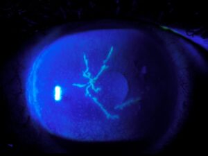
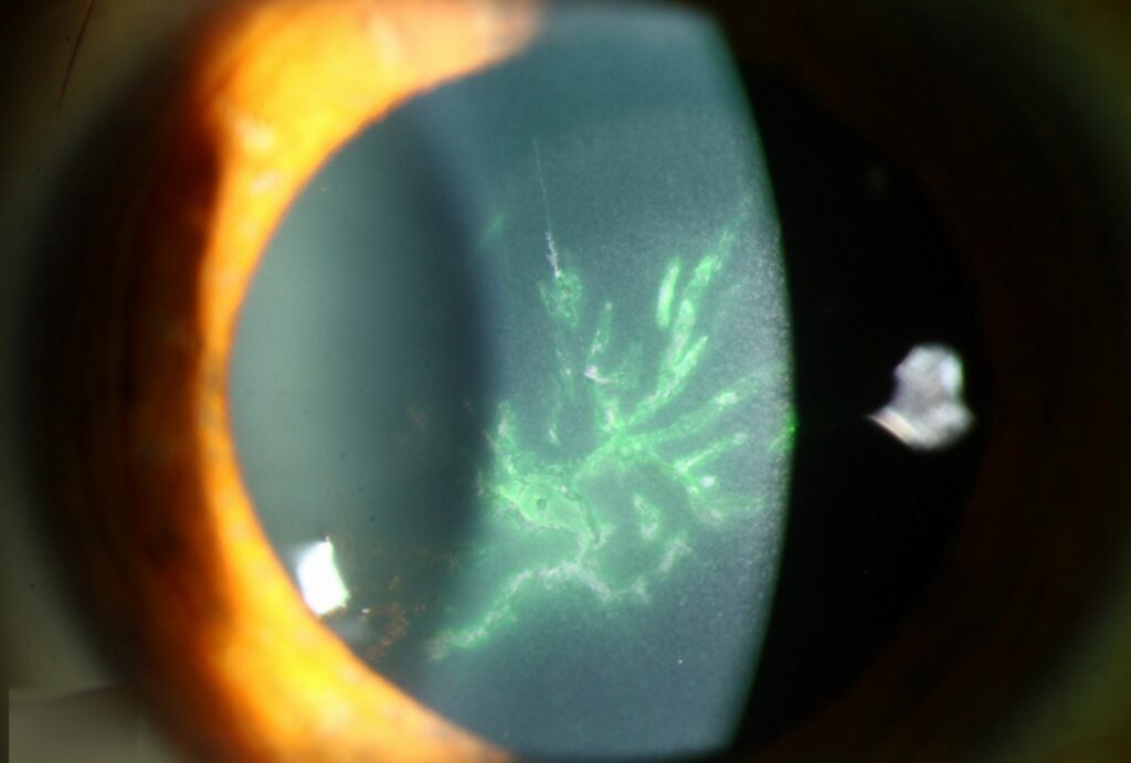
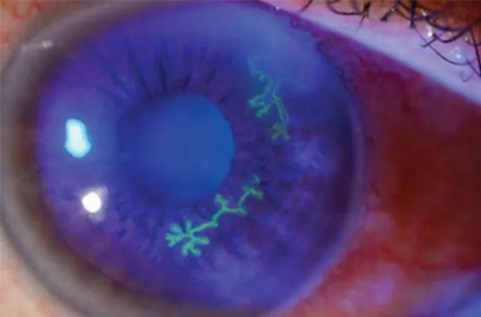
Infectious epithelial disease starts as punctate vesicles that coalesce into a branching dendrite. The branches end in terminal bulbs, and the ulcer bed stains with fluorescein, while the swollen margins take up rose bengal because they hold infected cells. In addition, corneal sensation is often reduced.
All-fit Slit-Lamp Adapter
Record and share exactly what you see at the slit lamp. One adapter fits any slit lamp or surgical microscope — and any smartphone.
From Choroida — the team behind this siteGeographic and Marginal Ulcers
A dendrite can enlarge into a geographic ulcer, especially after topical steroid use, and its scalloped edges still show dendritic tails (see geographic herpetic ulcers).
Marginal ulcers sit near the limbus and can mimic staphylococcal marginal keratitis. However, they usually show an overlying epithelial defect and adjacent limbal injection.
Treating Epithelial HSV Keratitis Types
First, start topical ganciclovir gel, trifluridine or an oral antiviral such as acyclovir. Oral therapy avoids the surface toxicity of frequent topical drops.
Gentle debridement of the dendrite can also lower the viral load. Then stop topical steroids where possible and review within a few days.
Neurotrophic Keratopathy After Herpes
Repeated episodes damage corneal nerves, so a smooth, rolled-edge defect can persist after the virus has cleared. Further antivirals only add toxicity, so treat the surface as neurotrophic disease.
Lubrication, a bandage lens or amniotic membrane can support healing.
Stromal Disease Among HSV Keratitis Types
Stromal keratitis causes much of the visual loss from herpes because it recurs and scars. However, the stroma shows two different patterns. One is driven by active virus, while the other is an immune reaction to viral antigen.
Necrotizing Stromal Keratitis
Necrotizing disease shows dense white infiltrates, stromal necrosis and often an epithelial defect, and thinning can progress quickly toward perforation.
Therefore, exclude bacterial or fungal infection before attributing it to herpes (see necrotizing stromal keratitis). Treatment combines antivirals with cautious steroids once the epithelium is stable.
Immune Stromal Keratitis
Immune stromal keratitis shows stromal haze, edema or ghost vessels under an intact epithelium, and an immune ring may surround the infiltrate.
The eye can look fairly quiet while vision falls from haze. Also, recurrent bouts add vascularization, lipid deposition and scarring.
Treating Stromal HSV Keratitis Types
In the Herpetic Eye Disease Study, topical prednisolone with trifluridine reduced persistence and progression of stromal keratitis. Deferring steroids did not worsen final visual acuity in that trial.
Therefore, start a topical steroid under oral antiviral cover, then taper slowly. Warn patients that rapid tapering often triggers rebound inflammation.
Endotheliitis and the Disciform Pattern
Endotheliitis causes stromal and epithelial edema overlying keratic precipitates. The disciform form gives a central round zone of edema, although diffuse and linear forms also occur.
The stroma shows no infiltrate, which separates it from immune stromal disease (see herpetic disciform keratitis).
Treating Endotheliitis
Treatment uses a topical steroid with oral antiviral cover. Meanwhile, check intraocular pressure because associated trabeculitis can raise it.
Sectoral iris atrophy also suggests herpetic uveitis, and persistent edema after repeated attacks usually reflects endothelial cell loss.
Overlap Between HSV Keratitis Types
Several layers can be active at once. For example, a dendrite may sit over immune stromal haze.
In that case, heal the epithelium first, then add steroids carefully, and also record each layer separately at every visit.
Diagnostic Tests for HSV Keratitis Types
Diagnosis is mainly clinical and relies on careful slit lamp examination. However, PCR of epithelial scrapings helps when the picture is atypical.
Test corneal sensation before instilling anesthetic drops. Then look for old scars and ghost vessels from earlier episodes.
Preventing Recurrence
The HEDS prevention trial found that long-term oral acyclovir reduced recurrent ocular herpes. The stromal benefit appeared in patients with prior stromal keratitis.
Therefore, offer prophylaxis to patients with frequent or vision-threatening attacks, and also explain that recurrences may return after stopping treatment.
Pitfalls to Avoid
- Do not give steroids alone for an active dendrite
- Never continue antivirals indefinitely for a neurotrophic defect
- Check pressure in every patient with endotheliitis
- Do not miss bacterial superinfection in a necrotic stroma
- Remember that recurrences often involve a different layer


Document what you see
Two smartphone imaging tools built for everyday clinic use — one for the slit lamp, one for the fundus.
From Choroida — the team behind this siteReferences
- Holland EJ, Schwartz GS. Classification of herpes simplex virus keratitis. Cornea. 1999;18:127-143.
- Wilhelmus KR, Gee L, Hauck WW, et al. Herpetic Eye Disease Study: a controlled trial of topical corticosteroids for herpes simplex stromal keratitis. Ophthalmology. 1994;101:1883-1896.
- Herpetic Eye Disease Study Group. Acyclovir for the prevention of recurrent herpes simplex virus eye disease. N Engl J Med. 1998;339:300-306.
- White ML, Chodosh J. Herpes simplex virus keratitis: a treatment guideline. American Academy of Ophthalmology; 2014.
- Bowling B. Kanski’s Clinical Ophthalmology: A Systematic Approach. 8th ed. Elsevier; 2016.
Test yourself
A few questions straight from this article.
-
The Holland and Schwartz scheme classifies HSV keratitis according to which feature?
The scheme sorts HSV keratitis by the corneal layer the virus or immune response involves, linking each sign to a mechanism and a first treatment step. -
The branches of an HSV epithelial dendrite characteristically end in which feature?
Punctate vesicles coalesce into a branching dendrite whose branches end in terminal bulbs, with a fluorescein-staining ulcer bed. -
How should immune-mediated HSV stromal keratitis be treated, as distinct from live viral replication?
Live virus needs antivirals, while immune disease needs steroids under antiviral cover, which is why the involved layer must be identified first. -
An HSV dendrite enlarging into a geographic ulcer is especially associated with which factor?
A dendrite can enlarge into a geographic ulcer especially after topical steroid use, and its scalloped edges still show dendritic tails. -
How should a smooth, rolled-edge defect persisting after HSV has cleared from the cornea be managed?
Repeated episodes damage corneal nerves; more antivirals only add toxicity, so lubrication, a bandage lens or amniotic membrane support healing. -
Which finding characterizes immune stromal HSV keratitis?
Immune stromal keratitis shows haze, edema or ghost vessels under an intact epithelium, sometimes with an immune ring around the infiltrate. -
Which feature separates HSV endotheliitis from immune stromal keratitis?
Endotheliitis causes stromal and epithelial edema over keratic precipitates, but the stroma shows no infiltrate. -
Why must intraocular pressure be checked in every patient with HSV endotheliitis?
Endotheliitis is treated with topical steroid under oral antiviral cover, and pressure is checked because associated trabeculitis can raise it. -
In the Herpetic Eye Disease Study, what effect did deferring topical steroids for stromal keratitis have on final acuity?
Topical prednisolone with trifluridine reduced persistence and progression of stromal keratitis, yet deferring steroids did not worsen final visual acuity. -
In the HEDS prevention trial, which treatment reduced recurrent ocular herpes?
Long-term oral acyclovir reduced recurrence, so prophylaxis is offered for frequent or vision-threatening attacks; recurrences may return after stopping.

Writes and reviews the clinical articles on Educate. Every post is checked against peer-reviewed sources before it goes live.