Dalen-Fuchs nodules are small yellow-white lesions at the level of the retinal pigment epithelium in eyes with granulomatous uveitis.
They are classic in sympathetic ophthalmia and Vogt-Koyanagi-Harada disease, and they also occur in sarcoidosis. Their presence points to a diffuse inflammatory process of the uvea and guides the workup.
What Dalen-Fuchs Nodules Are
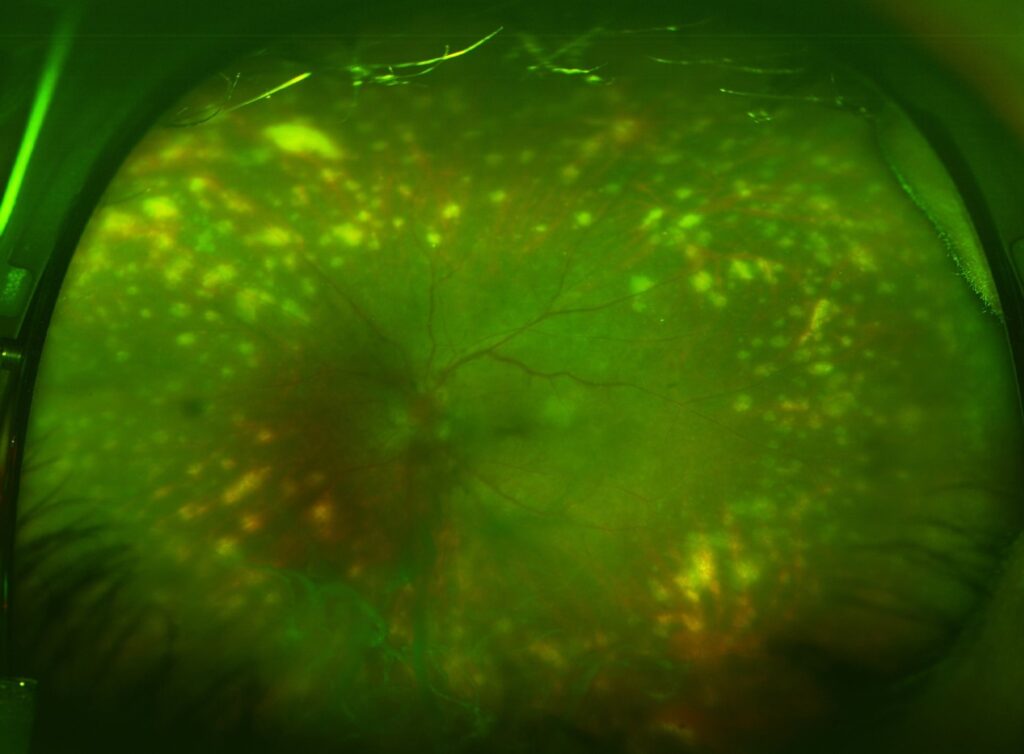
Histology shows collections of epithelioid cells, lymphocytes, and proliferating RPE cells between the RPE and Bruch membrane. Clinically they appear as multiple small, round, cream-colored spots in the mid-periphery.
Chan and colleagues described the immunopathology of sympathetic ophthalmia, including these lesions (see sympathetic ophthalmia). They do not cause symptoms by themselves, but they reflect active choroidal inflammation.
Dalen-Fuchs Nodules in Sympathetic Ophthalmia
Sympathetic ophthalmia is a bilateral granulomatous panuveitis after penetrating injury or surgery to one eye. The nodules are common in the fellow eye, and they appear along with vitritis and choroidal thickening. Damico and colleagues reviewed the disease and stressed the importance of early immunosuppression.
All-fit Slit-Lamp Adapter
Record and share exactly what you see at the slit lamp. One adapter fits any slit lamp or surgical microscope — and any smartphone.
From Choroida — the team behind this siteDalen-Fuchs Nodules in VKH Disease
In VKH disease, the nodules may appear in the late stage, along with sunset glow fundus and depigmentation. Read and colleagues reported that recurrent disease and complications correlate with chronic stages. Their presence helps to distinguish VKH from other causes of exudative detachment (see VKH disease and imaging).
Other Causes of Dalen-Fuchs Nodules
- Sarcoidosis and tuberculosis
- Lens-induced and other granulomatous uveitis
- Birdshot and other choroidopathies, in occasional reports
Look for systemic clues, such as hilar adenopathy or skin findings (see ocular sarcoidosis).
Imaging of Dalen-Fuchs Nodules
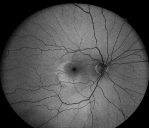
OCT shows small dome-shaped elevations of the RPE with a hyperreflective core, and fluorescein angiography shows hypofluorescence early and hyperfluorescence later, or a pinpoint leak. Indocyanine green angiography shows hypofluorescent spots that reflect choroidal granulomas, while autofluorescence shows spots with increased or decreased signal.
Management
Treat the underlying disease with high-dose corticosteroids and immunosuppressive agents, because sympathetic ophthalmia and VKH require early and sustained therapy to prevent recurrences. Evaluate for systemic causes, and monitor for complications such as glaucoma, cataract, and neovascularization.
An Example Case
A 38-year-old man had a penetrating injury to his right eye six weeks ago, and the left eye now shows photophobia and blurred vision. Examination shows mutton-fat keratic precipitates, vitritis, and multiple small yellow-white nodules in the mid-periphery.
OCT shows choroidal thickening and subretinal fluid, and angiography shows multiple pinpoint leaks. The diagnosis is sympathetic ophthalmia, and he receives high-dose corticosteroids and an immunosuppressive agent. The nodules fade over months, and his vision returns to 20/25.
Questions Patients Ask
Patients ask whether the nodules will go away, and they usually fade with treatment. They also ask whether the disease will return, and recurrences are possible without long-term control, so explain the need for lasting treatment because the disease affects both eyes.
Pitfalls to Avoid
- Mistaking the nodules for drusen or drusenoid deposits
- Stopping steroids too early in sympathetic ophthalmia or VKH
- Missing a history of ocular trauma or surgery
- Overlooking systemic causes such as sarcoidosis and tuberculosis
Looking Ahead
Wide-field imaging and OCT angiography may reveal nodules and choroidal inflammation more clearly, and biologic drugs may improve control with fewer steroid effects. Therefore, outcomes may continue to improve.
Practical Points
Ask about trauma, surgery, and systemic symptoms in every patient with granulomatous uveitis. Then image the choroid, order tests for sarcoidosis and tuberculosis when needed, and coordinate with a uveitis specialist early.
Distinguishing Nodules From Drusen
Drusen sit in older patients, and they have smooth, round shapes with no inflammation. Dalen-Fuchs nodules appear with active uveitis and in younger patients, so review the clinical context carefully because the treatment differs.
Histology in Brief
Biopsy is rarely needed, but pathology reveals clusters of epithelioid cells and pigmented cells under the RPE. Therefore, the nodules reflect a granulomatous process, and T-cell responses against retinal antigens drive the disease in sympathetic ophthalmia and VKH.
Course and Prognosis
With early and sustained treatment, most patients keep good vision. However, delayed treatment leads to cataract, glaucoma, and depigmentation, and because relapses may occur, follow patients over the years.
Counselling
Explain that the nodules themselves are not dangerous, but they signal active inflammation. Then discuss the plan for steroid tapering and drug monitoring.
Differential Diagnosis of Dalen-Fuchs Nodules
Drusen, multifocal choroiditis, and birdshot lesions may look similar. However, the history, the age, and the signs of inflammation separate them, so also use indocyanine green angiography to find choroidal granulomas. Therefore, a careful workup prevents a missed diagnosis, and because the diagnosis changes treatment, document the findings in detail.


Document what you see
Two smartphone imaging tools built for everyday clinic use — one for the slit lamp, one for the fundus.
From Choroida — the team behind this siteReferences
- Chan CC, Nussenblatt RB, Fujikawa LS, et al. Sympathetic ophthalmia: immunopathological findings. Ophthalmology. 1986;93:690-695.
- Damico FM, Kiss S, Young LH. Sympathetic ophthalmia. Semin Ophthalmol. 2005;20:191-197.
- Read RW, Rechodouni A, Butani N, et al. Complications and prognostic factors in Vogt-Koyanagi-Harada disease. Am J Ophthalmol. 2001;131:599-606.
- American Academy of Ophthalmology. Basic and Clinical Science Course, Section 9: Intraocular Inflammation and Uveitis. AAO; current edition.

Writes and reviews the clinical articles on Educate. Every post is checked against peer-reviewed sources before it goes live.