Hyphema recovery follows a fairly predictable course once the bleeding in the anterior chamber stops.
Most small traumatic hyphemas clear within about one week. However, the first five days carry the highest risk of rebleeding and pressure spikes, so patients need clear advice on rest, positioning and follow-up during that window.
How the Eye Clears the Blood
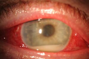
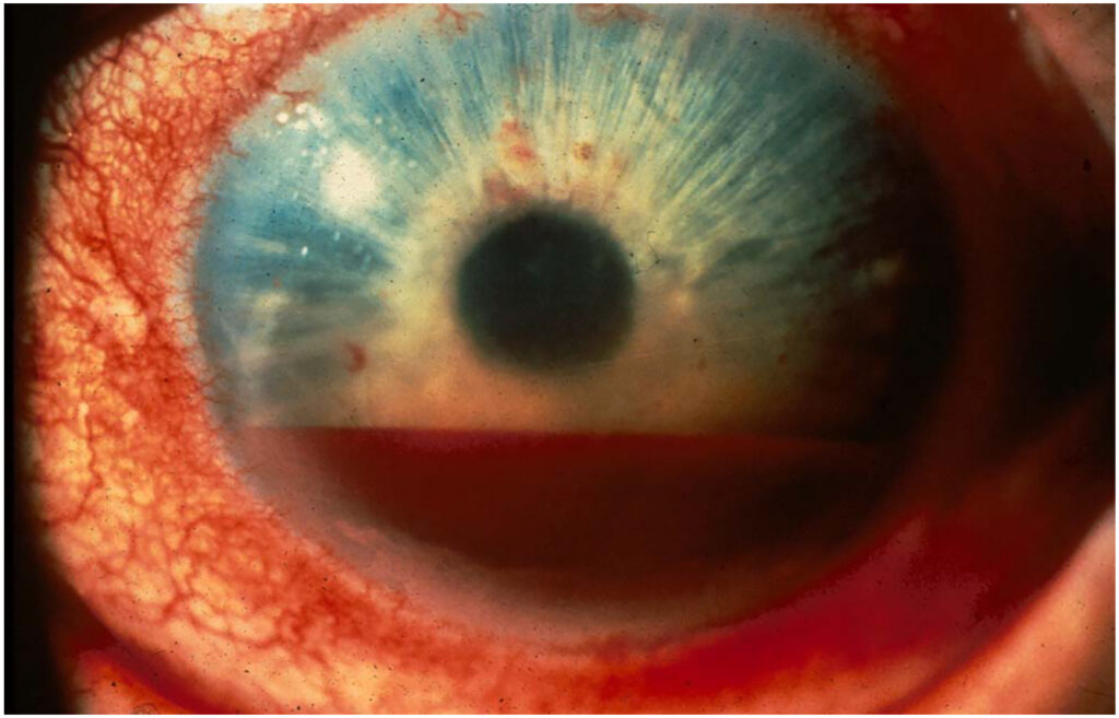
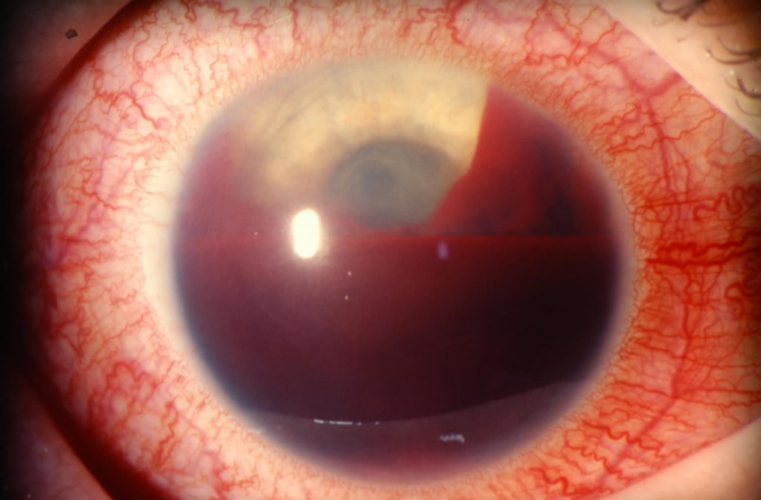
Blood in the anterior chamber settles inferiorly with gravity and forms a fluid level, while red cells leave the eye mainly through the trabecular meshwork. As a result, the level drops a little each day in an uncomplicated case.
Days 1 to 2 of Hyphema Recovery
On day one, the patient often reports a red haze or a dark shade in the lower field. The examiner measures the height of the layered blood in millimeters and checks pressure, and also notes circulating red cells, because they blur vision more than the layer itself. This baseline grade guides every later comparison (see hyphema grading).
All-fit Slit-Lamp Adapter
Record and share exactly what you see at the slit lamp. One adapter fits any slit lamp or surgical microscope — and any smartphone.
From Choroida — the team behind this siteDays 3 to 5: The Rebleed Window
Secondary hemorrhage occurs most often between the second and fifth day after injury, when the clot retracts and lyses, so the damaged vessel can bleed again. A rebleed is often larger than the first bleed and carries a worse visual outlook. Therefore, patients must report sudden blur, new pain or vomiting at once (see hyphema rebleed).
Day 6 Onward in Hyphema Recovery
In most uncomplicated eyes the level falls steadily and the clot shrinks. Microhyphemas often clear within a few days, while layered hyphemas usually take longer. However, clotted or total hyphemas may persist for weeks and need closer review.
Head Position and Shield
Ask the patient to sleep with the head raised about 30 to 45 degrees, which lets the blood settle below the visual axis. Meanwhile, a rigid shield protects the eye from accidental pressure or rubbing.
Activity Limits During Hyphema Recovery
Quiet activity at home suits most patients, and available trials comparing strict bed rest with moderate ambulation have not shown a clear advantage for bed rest. However, patients should avoid sports, heavy lifting and straining until the surgeon clears them.
Drops and Pain Relief
Topical corticosteroids reduce anterior chamber inflammation and improve comfort, while cycloplegic drops relieve ciliary spasm and may limit movement of the iris. Patients should avoid aspirin and NSAIDs, because they impair platelet function. Instead, acetaminophen usually controls pain without adding bleeding risk.
Pressure Spikes in Hyphema Recovery
Pressure may rise early from red cells blocking the meshwork, and later it may rise again with a rebleed or a clot that blocks the pupil. Check pressure at every visit during the first week. First-line treatment uses topical aqueous suppressants, such as beta-blockers.
Sickle Cell Risk in Hyphema Recovery
Sickled red cells pass through the meshwork poorly and raise pressure quickly, and the optic nerve in these patients tolerates even moderate pressure poorly. Therefore, screen at-risk patients for sickle cell disease or trait at presentation. Many authors advise avoiding carbonic anhydrase inhibitors, because acidosis may increase sickling.
When Surgery Becomes Necessary
Commonly cited thresholds for washout include high pressure that persists despite treatment. For example, Read and Goldberg proposed surgery for pressure of 60 mmHg or more for two days. Early corneal blood staining and long-lasting total hyphema are also indications.
When Vision Should Return
Vision usually improves as the blood leaves the visual axis, which in small hyphemas often happens within the first week. If vision stays poor after the chamber clears, look for associated injury:
- Traumatic cataract or lens subluxation
- Commotio retinae or choroidal rupture
- Traumatic optic neuropathy
- Corneal blood staining (see corneal blood staining)
Corneal Staining in Hyphema Recovery
Staining develops with high pressure, prolonged blood contact and endothelial damage, and the stained cornea turns brown and blurs the view of the iris. It clears slowly from the periphery toward the center over many months, and in young children this delay can cause amblyopia.
Follow-Up After the Blood Clears
Perform gonioscopy once the chamber is clear, often a few weeks after injury, and look for angle recession, which raises the long-term risk of glaucoma. Next, examine the dilated fundus for retinal tears and dialysis. Patients with angle recession then need lifelong yearly pressure checks.
Pitfalls to Avoid
- Do not stop pressure checks once the blood level falls
- Never prescribe aspirin or NSAIDs for pain after hyphema
- Do not miss sickle cell status in at-risk patients
- Avoid scleral depression during the rebleed window


Document what you see
Two smartphone imaging tools built for everyday clinic use — one for the slit lamp, one for the fundus.
From Choroida — the team behind this siteReferences
- Walton W, Von Hagen S, Grigorian R, Zarbin M. Management of traumatic hyphema. Surv Ophthalmol. 2002;47:297-334.
- Gharaibeh A, Savage HI, Scherer RW, Goldberg MF, Lindsley K. Medical interventions for traumatic hyphema. Cochrane Database Syst Rev. 2019;1:CD005431.
- Read J, Goldberg MF. Comparison of medical treatment for traumatic hyphema. Trans Am Acad Ophthalmol Otolaryngol. 1974;78:799-815.
- Bowling B. Kanski’s Clinical Ophthalmology: A Systematic Approach. 8th ed. Edinburgh: Elsevier; 2016.

Writes and reviews the clinical articles on Educate. Every post is checked against peer-reviewed sources before it goes live.