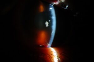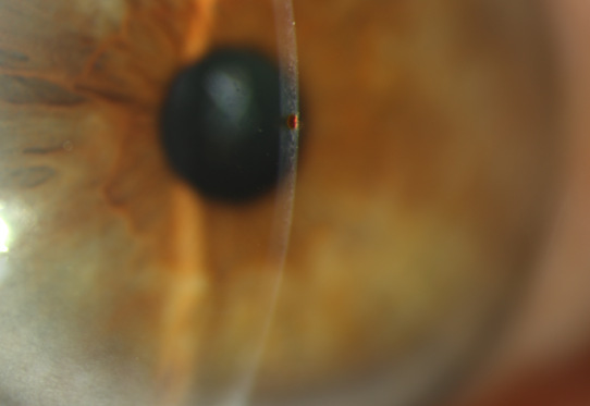
Subtarsal foreign body describes a particle trapped on the tarsal conjunctiva under the upper lid.
The patient usually reports a scratchy eye that worsens with every blink.
However, the particle stays hidden until the examiner everts the lid.
Where a Subtarsal Foreign Body Lodges
Particles often settle in the subtarsal sulcus, a shallow groove near the upper lid margin.
Then the tarsal plate holds the particle against the moving cornea.
As a result, each blink drags it across the epithelium.
Symptoms of a Subtarsal Foreign Body
Patients describe grittiness, tearing, photophobia and sharp discomfort on blinking.
Vision may stay normal, or it may blur when scratches cross the visual axis.
In addition, symptoms often begin suddenly, for example after wind or a workshop task.
All-fit Slit-Lamp Adapter
Record and share exactly what you see at the slit lamp. One adapter fits any slit lamp or surgical microscope — and any smartphone.
From Choroida — the team behind this siteHistory That Shapes the Exam
First, ask exactly what the patient was doing when symptoms began.
Hammering metal and grinding raise concern for a high-velocity fragment.
Next, ask about contact lens wear and previous eye surgery.
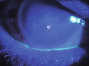
Vertical Staining With a Subtarsal Foreign Body
Fluorescein often reveals fine, parallel, vertical lines on the upper cornea.
These lines appear because the blink moves the particle over the epithelium.
Therefore, linear vertical staining should prompt lid eversion in every case (see corneal staining patterns).
Slit Lamp Survey Before Eversion
Begin with visual acuity, then inspect the lids, conjunctiva, cornea and anterior chamber.
Look for infiltrate, hyphema, an irregular pupil or a conjunctival laceration.
If you suspect a globe injury, avoid pressure and defer eversion.
Everting the Lid for a Subtarsal Foreign Body
A drop of topical anesthetic reduces blepharospasm.
Next, ask the patient to look down and keep looking down.
Grasp the upper lashes, then press the tarsal border gently with a cotton-tipped applicator.
Finally, rotate the lid over the applicator and inspect it under magnification (see eyelid eversion technique).
Searching the Superior Fornix
Single eversion shows the tarsal conjunctiva, while the fornix stays out of view.
Therefore, persistent symptoms after a negative exam call for double eversion with a Desmarres retractor.
Alternatively, a moistened applicator can sweep the fornix after anesthesia.
Removing a Subtarsal Foreign Body
Most particles lift away easily with a moistened cotton-tipped applicator.
Irrigation also helps when several loose particles coat the conjunctiva.
An embedded fragment may need fine forceps or a needle at the slit lamp.
However, avoid repeated rubbing, because it can abrade the conjunctiva.
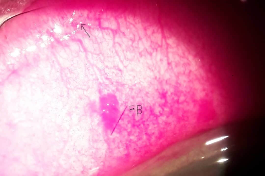
Checking for Residual Damage
After removal, stain the cornea again and record the size and position of any defect.
Then look for additional particles, because windblown exposure can leave several fragments.
Symptoms usually improve quickly, although the defect may stay tender until it heals.
Excluding Penetrating Injury
A subtarsal particle does not exclude a second fragment inside the eye.
High-velocity mechanisms therefore need a Seidel test and a dilated fundus examination when safe.
In addition, order CT if a metallic intraocular fragment remains possible (see intraocular foreign body).
Treating the Corneal Scratches
Small abrasions commonly receive a topical antibiotic for a short course.
Lubricants also improve comfort, while topical NSAIDs can reduce pain.
A Cochrane review found that patching did not speed healing in simple abrasions.
However, patients still need clear return instructions.
Contact Lens Wearers
Abrasions in lens wearers carry a higher risk of bacterial keratitis.
Therefore, stop lens wear and choose antipseudomonal coverage, for example a fluoroquinolone.
Also, check the fornix carefully when a patient cannot account for a lost lens.
Differential Diagnosis of Subtarsal Foreign Body
Conjunctival concretions can produce a similar scratchy sensation, and they also appear on eversion.
Trichiasis, loose sutures and giant papillary conjunctivitis may also abrade the upper cornea.
Meanwhile, herpes simplex keratitis deserves attention when dendritic staining appears.
Follow-Up and Counseling
Small, clean abrasions in reliable patients may not need a routine recheck.
However, review larger or central defects and contact lens wearers within a day or two.
An infiltrate, worsening pain or falling acuity calls for urgent reassessment.
Finally, advise protective eyewear and avoid home topical anesthetic, because repeated use delays healing.
Pitfalls to Avoid
- Do not treat vertical staining as dry eye before everting the lid
- Never stop searching after finding one particle
- Avoid eversion when an open globe seems likely
- Skipping a dilated exam after a high-velocity injury is a mistake
- Never patch an abrasion in a contact lens wearer


Document what you see
Two smartphone imaging tools built for everyday clinic use — one for the slit lamp, one for the fundus.
From Choroida — the team behind this siteReferences
- Wipperman JL, Dorsch JN. Evaluation and management of corneal abrasions. Am Fam Physician. 2013;87:114-120.
- Lim CH, Turner A, Lim BX. Patching for corneal abrasion. Cochrane Database Syst Rev. 2016;7:CD004764.
- Bagheri N, Wajda BN, eds. The Wills Eye Manual. 7th ed. Wolters Kluwer; 2017.
- Bowling B. Kanski’s Clinical Ophthalmology. 8th ed. Elsevier; 2016.
- American Academy of Ophthalmology. Basic and Clinical Science Course, Section 8: External Disease and Cornea. AAO; latest edition.

Writes and reviews the clinical articles on Educate. Every post is checked against peer-reviewed sources before it goes live.