
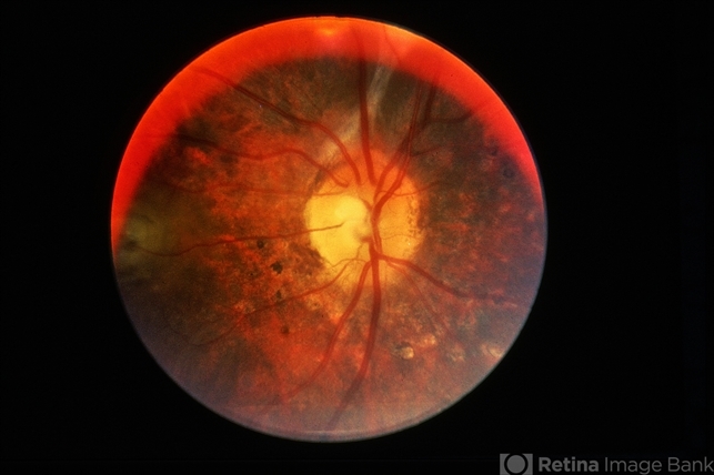
Vogt–Koyanagi–Harada (VKH) at a glance
- Also known as: Vogt–Koyanagi–Harada syndrome, uveomeningeal syndrome
- ICD-10: H20.82 (Vogt–Koyanagi syndrome) · H30.81 (Harada’s disease)
- Core lesion: Bilateral granulomatous panuveitis driven by choroidal inflammation
- Key association: HLA-DRB1*04:05; higher prevalence in Asian, Hispanic, Middle Eastern, and Native American populations
- Most useful activity biomarker: Choroidal thickness on EDI/SS-OCT and hypofluorescent dark dots on ICGA
Key Takeaways
- VKH is fundamentally a disease of the choroid; retinal findings are secondary to choroidal inflammation.
- Multimodal imaging (EDI-OCT, SS-OCT, ICGA, OCTA, FAF, ultra-widefield) now detects subclinical activity that clinical exam and fluorescein angiography can miss.
- Choroidal thickness has become a practical, quantifiable biomarker of disease activity and treatment response.
- ICGA is among the most sensitive tools for identifying persistent, subclinical inflammation.
- Persistent imaging abnormalities can justify continued immunosuppression even after vision improves.
Vogt–Koyanagi–Harada (VKH) disease is a multisystem autoimmune disorder characterized by bilateral granulomatous panuveitis, with inflammation directed against melanocyte-containing tissues.[4] Although VKH has long been recognized as a major cause of inflammatory serous retinal detachment and vision loss, our understanding of the disease has changed dramatically over the past two decades.
Traditionally, diagnosis relied heavily on clinical findings and fluorescein angiography. Today, advances in multimodal imaging—including enhanced-depth imaging OCT (EDI-OCT), swept-source OCT (SS-OCT), OCT angiography (OCTA), indocyanine green angiography (ICGA), and ultra-widefield imaging—have transformed the diagnosis, monitoring, and management of VKH.[6]
Modern imaging has shown that VKH is fundamentally a disease of the choroid, with retinal findings representing secondary consequences of choroidal inflammation.[3] For ophthalmologists, mastering these modalities has become essential for detecting subclinical activity, guiding treatment, and preventing chronic complications.
Understanding VKH Disease
VKH is an autoimmune disorder in which T-cell–mediated inflammation targets melanocytes within the eye, meninges, inner ear, skin, and hair.[4] The disease typically progresses through four clinical stages:
- Prodromal stage
- Acute uveitic stage
- Convalescent stage
- Chronic recurrent stage
Although patients often present during the acute phase, imaging can detect inflammatory changes before irreversible structural damage develops.
Modern imaging has shifted the focus from retinal pathology to choroidal inflammation.
Revised Diagnostic Criteria (2001)
The internationally accepted Revised Diagnostic Criteria classify VKH by the extent of extraocular involvement, after excluding prior ocular trauma/surgery and other ocular disease.[1]
| Category | Required features |
|---|---|
| Complete VKH | No prior penetrating trauma/surgery · no other ocular disease · bilateral ocular involvement · plus neurological/auditory findings · plus integumentary findings (poliosis, vitiligo, alopecia) |
| Incomplete VKH | Bilateral ocular involvement plus either neurological/auditory or integumentary findings |
| Probable VKH | Isolated bilateral ocular disease, with no neurological, auditory, or skin findings |
A history of penetrating ocular trauma or surgery excludes VKH and should redirect suspicion toward sympathetic ophthalmia.
Fundus Explorer Pro
Photograph the retinal findings described here with the phone already in your pocket — 22 D optics and built-in illumination in one handheld unit.
From Choroida — the team behind this siteWhy Modern Imaging Changed VKH Management
Historically, clinicians relied mainly on clinical examination and fluorescein angiography (FA). While still valuable, these methods do not always detect early or persistent choroidal inflammation. Today’s imaging allows clinicians to detect disease earlier, monitor treatment response objectively, identify subclinical recurrence, tailor immunosuppressive therapy, and predict visual prognosis.
Imaging now guides treatment decisions as much as clinical examination.
Imaging modalities at a glance
| Modality | Key VKH finding / role |
|---|---|
| EDI-OCT / SS-OCT | Choroidal thickening, subretinal septa, RPE undulation; quantifies activity via choroidal thickness |
| Fluorescein angiography | “Starry-sky” pinpoint leaks, pooling in serous detachments, disc leakage |
| ICGA | Hypofluorescent dark dots; most sensitive for subclinical choroidal inflammation |
| OCTA | Choriocapillaris flow deficits; non-invasive vascular monitoring |
| FAF | Documents RPE damage and chronic pigmentary change |
| Ultra-widefield | Reveals peripheral choroidal/retinal involvement missed on standard photos |
Enhanced-Depth Imaging OCT (EDI-OCT)
One of the most important advances in VKH has been the ability to visualize the choroid in vivo.[3]
Acute VKH Findings
EDI-OCT commonly demonstrates marked choroidal thickening, serous retinal detachment, choroidal folds, undulating retinal pigment epithelium (RPE), and subretinal septa. These findings reflect intense inflammation and exudation within the choroid.
During Treatment
Successful therapy typically results in a progressive reduction in choroidal thickness, resolution of subretinal fluid, and restoration of foveal contour.
Choroidal thickness has become an important biomarker for disease activity.
Swept-Source OCT (SS-OCT)
Swept-source OCT provides deeper penetration and superior visualization of the choroid than conventional OCT, including better imaging of the thickened choroid, clearer view of the choroid–scleral interface, more accurate choroidal thickness measurements, and enhanced follow-up of chronic disease. SS-OCT is particularly useful for monitoring patients on long-term immunosuppressive therapy.
OCT Findings in Acute VKH
Several retinal abnormalities are characteristic of acute VKH: multilobular serous retinal detachment, intraretinal cystoid spaces, subretinal septa, RPE undulations, and hyperreflective dots within subretinal fluid. Among these, multilobular serous detachments are considered highly characteristic.
Subretinal septa are one of the classic OCT features of acute VKH.
Fluorescein Angiography (FA)
Despite newer technologies, FA remains an important diagnostic tool. Early-phase findings include delayed choroidal perfusion and multiple pinpoint hyperfluorescent leaks; late-phase findings include pooling within serous detachments, diffuse optic disc leakage, and progressive dye accumulation. The “starry-sky” appearance produced by numerous pinpoint leaks remains a classic angiographic hallmark. For a broader review of leakage patterns, see leaky vessels on angiography.
Indocyanine Green Angiography (ICGA)
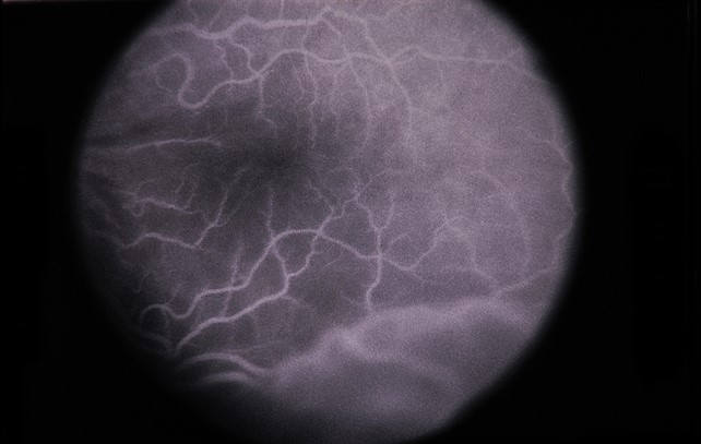
ICGA has transformed the evaluation of choroidal inflammation because, unlike fluorescein angiography, it directly assesses the choroidal circulation.[2] Typical findings include multiple hypofluorescent dark dots, choroidal stromal inflammation, fuzzy choroidal vessels, and delayed choroidal perfusion. ICGA frequently detects persistent inflammation even after clinical improvement.[5]
ICGA is one of the most sensitive methods for identifying subclinical VKH activity.
OCT Angiography (OCTA)
Although OCTA does not demonstrate leakage, it provides valuable information about retinal and choroidal microvasculature, including areas of choriocapillaris flow deficit, reduced vascular density, and microvascular remodeling during recovery. OCTA complements conventional angiography with a non-invasive vascular assessment.
Fundus Autofluorescence (FAF)
FAF helps evaluate retinal pigment epithelium integrity. In acute disease it may show mixed hyperautofluorescence and areas corresponding to serous detachments; in chronic disease it can reveal RPE atrophy, hypoautofluorescent scars, and chronic pigmentary changes. FAF is particularly useful for documenting long-term structural damage.
Ultra-Widefield Imaging
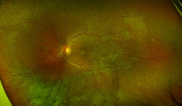
Peripheral inflammation may be underestimated on conventional fundus photography. Ultra-widefield imaging allows visualization of peripheral choroidal inflammation, peripheral retinal involvement, and widespread pigmentary alterations—improving disease monitoring in chronic VKH.
Imaging Features by Disease Stage
Acute Uveitic Stage
Choroidal thickening, serous retinal detachments, pinpoint FA leakage, and hypofluorescent ICGA lesions.
Convalescent Stage
Resolution of subretinal fluid, decreasing choroidal thickness, and early pigmentary changes.
Chronic Recurrent Stage
Sunset-glow fundus, choroidal thinning, RPE atrophy, and recurrent inflammatory lesions.
Imaging findings evolve considerably throughout the course of VKH.
Modern Imaging Biomarkers of Disease Activity
Several imaging biomarkers help identify active inflammation: increased choroidal thickness, new serous retinal detachment, persistent hypofluorescent ICGA lesions, new pinpoint FA leakage, and choriocapillaris flow deficits on OCTA. These biomarkers often detect relapse before visual symptoms develop.
Differential Diagnosis
Because VKH shares features with several causes of bilateral serous detachment and panuveitis, imaging and history are used together to distinguish it.
| Condition | Key clue that argues against VKH |
|---|---|
| Sympathetic ophthalmia | History of penetrating ocular trauma or surgery to the fellow eye (imaging can be near-identical) |
| Central serous chorioretinopathy | Non-inflammatory; focal RPE leak; steroids worsen rather than help |
| Posterior scleritis | Pain, scleral thickening and “T-sign” on B-scan; often unilateral |
| Sarcoidosis | Systemic signs, hilar lymphadenopathy, elevated ACE, non-caseating granulomas |
| Uveal effusion / bullous exudative RD | Scleral thickening or nanophthalmos; no CNS, auditory, or skin findings |
How Imaging Guides Treatment
Imaging now plays a central role in treatment decisions. At initial evaluation it helps confirm the diagnosis, assess disease severity, and establish baseline anatomy. During follow-up it helps determine response to corticosteroids, the need for immunosuppressive therapy, the risk of recurrence, and the timing of treatment taper.
Persistent imaging abnormalities may justify continued immunosuppression even when vision has improved.
Future Perspectives
Emerging technologies continue to improve VKH management. Areas of ongoing research include artificial-intelligence–assisted image interpretation, quantitative choroidal biomarkers, automated inflammation scoring, widefield OCT, and molecular imaging. These advances may allow even earlier detection of disease activity and more individualized treatment.
Clinical Pearls
- Think of VKH as a choroidal disease first; the retina suffers secondarily.
- Choroidal thickness is a fast, repeatable activity biomarker—track it at every visit.
- ICGA and OCT can flag subclinical inflammation; do not taper on visual acuity alone.
- Always exclude penetrating trauma/surgery before diagnosing VKH—otherwise consider sympathetic ophthalmia.
Frequently Asked Questions
What is Vogt–Koyanagi–Harada disease?
VKH is a multisystem autoimmune disorder in which T-cell–mediated inflammation targets melanocytes in the eye, meninges, inner ear, skin, and hair, producing a bilateral granulomatous panuveitis.
Is VKH a disease of the retina or the choroid?
Primarily the choroid. Modern imaging shows that retinal findings such as serous detachments are secondary to diffuse choroidal inflammation.
Which imaging test is most sensitive for subclinical VKH activity?
ICGA is among the most sensitive tools for detecting persistent choroidal inflammation, complemented by choroidal thickness measurement on EDI-OCT or SS-OCT.
How is VKH diagnosed?
Using the Revised Diagnostic Criteria (2001): bilateral ocular involvement plus neurological/auditory and/or integumentary findings, after excluding prior ocular trauma or surgery and other ocular disease.
How is VKH distinguished from sympathetic ophthalmia?
Imaging findings can be nearly identical; the key differentiator is a history of penetrating ocular trauma or surgery, which points to sympathetic ophthalmia rather than VKH.


Document what you see
Two smartphone imaging tools built for everyday clinic use — one for the slit lamp, one for the fundus.
From Choroida — the team behind this siteReferences
- Read RW, et al. “Revised diagnostic criteria for Vogt-Koyanagi-Harada disease: report of an international committee on nomenclature.” Am J Ophthalmol. 2001. [PubMed]
- Herbort CP Jr, et al. “Indocyanine green angiography in Vogt-Koyanagi-Harada disease.” Int Ophthalmol. 2007. [PubMed]
- Chee SP, et al. “Enhanced depth imaging optical coherence tomography in Vogt-Koyanagi-Harada disease.” Ophthalmology. 2013. [PubMed]
- Yang P, et al. “Clinical characteristics and imaging findings in Vogt-Koyanagi-Harada disease.” Prog Retin Eye Res. 2018. [PubMed]
- Bouchenaki N, Herbort CP Jr. “Indocyanine green angiography-guided management of Vogt-Koyanagi-Harada disease.” Int Ophthalmol. 2011. [PubMed]
- Da Silva FT, et al. “Multimodal imaging in Vogt-Koyanagi-Harada disease.” Ocul Immunol Inflamm. 2016. [PubMed]
Test yourself
A few questions straight from this article.
-
In Vogt-Koyanagi-Harada disease, T-cell-mediated inflammation is directed against which cells?
VKH is a multisystem autoimmune disorder in which T-cell-mediated inflammation targets melanocyte-containing tissues of the eye, meninges, inner ear, skin and hair. -
Modern imaging shows Vogt-Koyanagi-Harada disease is fundamentally a disease of which structure?
Multimodal imaging established that VKH is primarily a choroidal disease, with retinal findings such as serous detachment arising secondarily from diffuse choroidal inflammation. -
Which HLA allele is the key genetic association of Vogt-Koyanagi-Harada disease?
VKH is associated with HLA-DRB1*04:05, and prevalence is higher in Asian, Hispanic, Middle Eastern and Native American populations. -
Under the Revised 2001 criteria, complete Vogt-Koyanagi-Harada disease requires which combination?
Complete VKH needs bilateral ocular involvement together with both neurological or auditory findings and integumentary findings such as poliosis, vitiligo or alopecia. -
A history of penetrating ocular trauma should redirect suspicion away from VKH toward which diagnosis?
Prior penetrating trauma or surgery excludes VKH under the diagnostic criteria; imaging in sympathetic ophthalmia can be near-identical, so the history is the differentiator. -
Which imaging test is most sensitive for subclinical choroidal inflammation in VKH disease?
ICGA directly assesses the choroidal circulation and shows hypofluorescent dark dots; it frequently detects persistent inflammation even after clinical improvement. -
Which quantifiable OCT measurement serves as an activity biomarker in Vogt-Koyanagi-Harada disease?
Choroidal thickness measured on enhanced-depth or swept-source OCT is a practical, repeatable biomarker; successful therapy produces a progressive reduction in thickness. -
Which fluorescein angiography appearance is the classic hallmark of acute VKH disease?
Numerous pinpoint hyperfluorescent leaks produce the classic starry-sky appearance, with late pooling in serous detachments and diffuse optic disc leakage. -
Which imaging picture characterises the chronic recurrent stage of Vogt-Koyanagi-Harada disease?
The chronic recurrent stage shows sunset-glow fundus, choroidal thinning, RPE atrophy and recurrent inflammatory lesions, in contrast to the thickened choroid of acute disease. -
In VKH disease, what may persistent imaging abnormalities justify once vision has improved?
Because ICGA and OCT can flag subclinical inflammation, persistent imaging abnormalities may justify continued immunosuppression; treatment should not be tapered on visual acuity alone.
