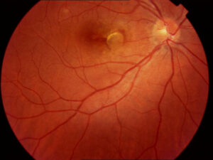
Amiodarone optic neuropathy is a rare toxic injury of the optic nerve in patients who take the antiarrhythmic drug amiodarone for months or years.
It is usually insidious and often bilateral.
The disc stays swollen for a long time, which makes it look like but behave differently from nonarteritic anterior ischemic optic neuropathy (see non-arteritic anterior ischemic optic neuropathy).
Because the drug can control life-threatening arrhythmias, the decision to stop it belongs to the cardiologist and the ophthalmologist together.
How Common Is Amiodarone Optic Neuropathy?
Estimates suggest an incidence of 1 to 2 percent of long-term users, though true rates are hard to know (see the references).
Most cases arise after months to years of therapy, with doses of 200 milligrams a day or more, although neuropathy has been seen at lower doses.
The condition is far less common than the corneal deposits that occur in almost all patients.
Amiodarone Optic Neuropathy: Eye Effects That Are Harmless
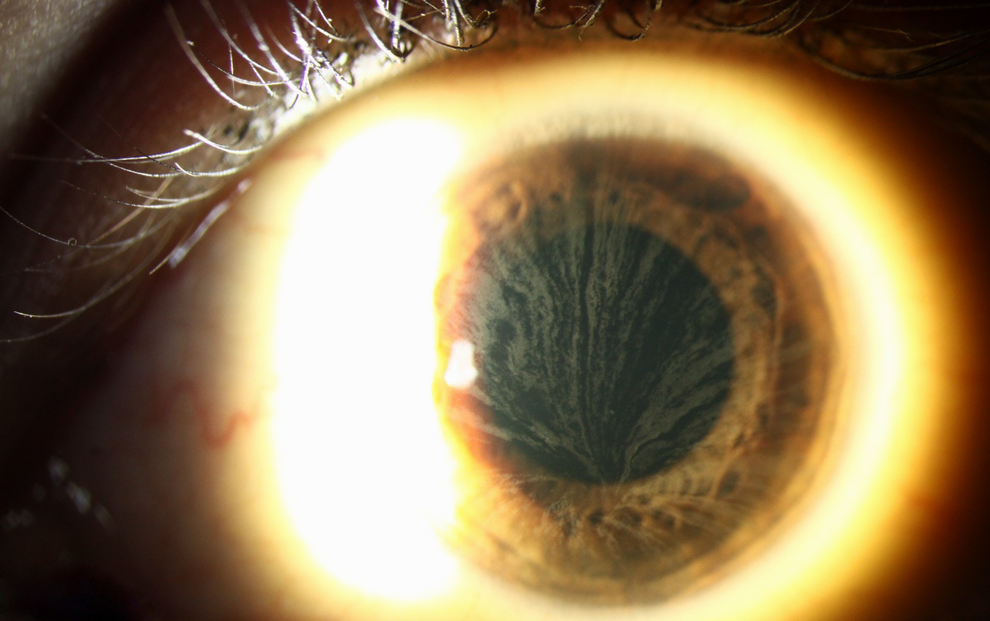
- Corneal verticillata: whorl-like deposits in the inferior cornea, seen in nearly every long-term user, which seldom affect vision (see cornea verticillata)
- Anterior subcapsular lens deposits: mild and rarely visually significant
These findings do not predict neuropathy, and they do not need treatment.
All-fit Slit-Lamp Adapter
Record and share exactly what you see at the slit lamp. One adapter fits any slit lamp or surgical microscope — and any smartphone.
From Choroida — the team behind this siteAmiodarone Optic Neuropathy: Clinical Features of the Neuropathy
Patients may notice mild blurring, or have no complaints at all, and routine examination may find the problem.
- Gradual, painless loss of acuity, often mild at first
- Visual field defects, such as enlarged blind spots, arcuate or central loss
- Bilateral disc swelling in most patients, with hyperemia and few hemorrhages
- A relative afferent pupillary defect if the loss is asymmetric
The disc swelling lasts for months, much longer than in NAION, and then resolves into pallor (see the references).
Differences From NAION
NAION is usually unilateral, abrupt, and has altitudinal loss with a crowded disc, and the swelling resolves in about two months.
Amiodarone neuropathy has an insidious onset, is more often bilateral, and has a longer swelling phase and a slower course.
Patients on amiodarone can also have NAION, so the distinction is not always clear.
It is a reason to review the whole history (see optic disc edema).
Evaluation for Amiodarone Optic Neuropathy
Take the drug history with dose and duration, and examine acuity, color, pupils, fields, and the disc.
Obtain OCT of the nerve fiber layer, and arrange MRI of the brain and orbits and blood tests when the picture is atypical, to exclude compression, inflammation, and other causes (see optic neuritis).
Management for Amiodarone Optic Neuropathy
Discuss the findings with the cardiologist, and decide whether the drug can be replaced.
If the cardiologist can stop it, the swelling usually resolves in two to six months.
Some patients regain vision, though many do not, and a few worsen after stopping (see the references).
If the drug must continue, the cardiologist may lower the dose, with close follow-up of the field and the disc.
Screening
There is no proven screening program.
Baseline eye examination before starting is reasonable, with periodic checks and a clear instruction to report visual changes.
Counselling
Explain that vision loss from this drug is rare, that it is a reason to call, and that no one should stop the medicine without medical advice.
An Example Case
A 68-year-old man with atrial fibrillation has taken amiodarone 200 milligrams daily for two years and complains that his vision “looks cloudy” in both eyes for about four months.
Acuity is 20/40 in each eye, both discs are swollen with little hemorrhage, there are enlarged blind spots, and the corneas show verticillata.
MRI is normal, and after discussion with his cardiologist, another agent replaces amiodarone.
The swelling resolves over five months, with acuity of 20/30.
Practical Steps in the Clinic
- Ask about the dose and duration in every patient with an unexplained swollen disc
- Examine both discs, and compare with old photographs if available
- Order the tests needed to exclude other causes, and do not label the case until the results return
- Write to the prescriber with clear findings, and give a recommendation
Counselling
Explain that most people who take this medicine never have eye problems, that the corneal changes are harmless, and that new blurring should be reported.
Reassure patients that the heart rhythm medicine is not stopped without a plan, and that the doctors will work together.
Key Points to Remember
- Insidious onset, often bilateral, with a long period of disc swelling
- Corneal verticillata are common and benign
- NAION can occur in patients on amiodarone, so keep the differential open
- Stopping the drug may lead to resolution of swelling, with variable recovery of vision
Limits of the Evidence
Reports are mainly case series and retrospective studies, and the true risk is hard to define.
Better data on dose, duration, and risk factors would help to guide screening.
For the latest studies, see the PubMed results for amiodarone optic neuropathy.


Document what you see
Two smartphone imaging tools built for everyday clinic use — one for the slit lamp, one for the fundus.
From Choroida — the team behind this siteReferences
- Macaluso DC, Shults WT, Fraunfelder FT. Features of amiodarone-induced optic neuropathy. Am J Ophthalmol. 1999;127:610-612.
- Purvin V, Kawasaki A, Borruat FX. Optic neuropathy in patients using amiodarone. Arch Ophthalmol. 2006;124:696-701.
- Passman RS, Bennett CL, Purpura JM, et al. Amiodarone-associated optic neuropathy: a critical review. Am J Med. 2012;125:447-453.
- American Academy of Ophthalmology. Basic and Clinical Science Course, Section 5: Neuro-Ophthalmology. AAO; current edition.

Writes and reviews the clinical articles on Educate. Every post is checked against peer-reviewed sources before it goes live.