
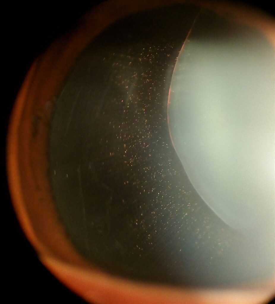
Ectopia lentis — displacement of the crystalline lens from its normal position — is one of the cardinal ocular features of Marfan syndrome, present in a large majority of affected individuals and, in many cases, the finding that first brings the underlying connective tissue disorder to medical attention.
The lens in Marfan syndrome typically subluxates upward and temporally, a pattern that is different enough from other causes of lens dislocation to be useful diagnostically.
Marfan syndrome is caused by mutations in FBN1, the gene encoding fibrillin-1, a major structural component of the zonular fibers that suspend the lens, and weakened or fragmented zonules under normal mechanical stress are what allow the lens to drift out of position, usually gradually rather than acutely.
Clinical Presentation
Many patients are diagnosed in childhood, because the zonular weakness is congenital even though the degree of visible subluxation often progresses over the first one to two decades of life.
Symptoms depend on the degree and direction of displacement: mild subluxation may cause only monocular diplopia or asymmetric astigmatism from the tilted or displaced lens edge, while more significant subluxation causes markedly reduced vision, often correctable only by finding the right optical zone or by aphakic correction if the lens has moved fully out of the visual axis.
Because the lens is typically off-axis rather than sinking posteriorly (as more often happens in traumatic dislocation), patients frequently describe a “second image” or monocular diplopia rather than sudden, profound vision loss.
Because this gradual, progressive pattern differs so much from the sudden onset typical of traumatic lens dislocation, a patient or family reporting slowly worsening double vision in one eye over months to years, rather than an acute event, should specifically raise suspicion for an underlying connective tissue disorder like Marfan syndrome.
Exam Findings
- Iridodonesis and phacodonesis — tremulousness of the iris and lens, respectively, on eye movement, from loss of normal zonular support
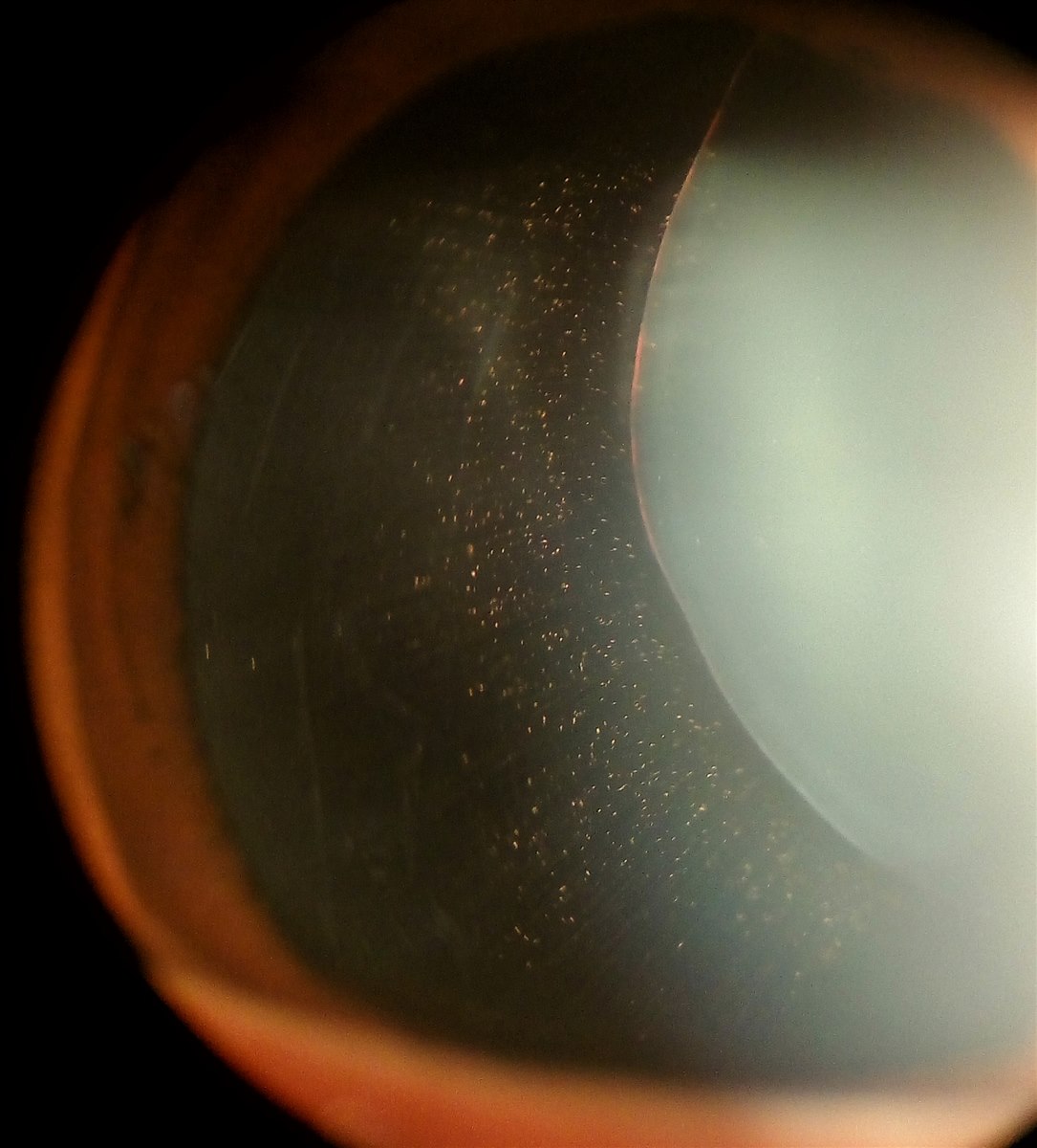
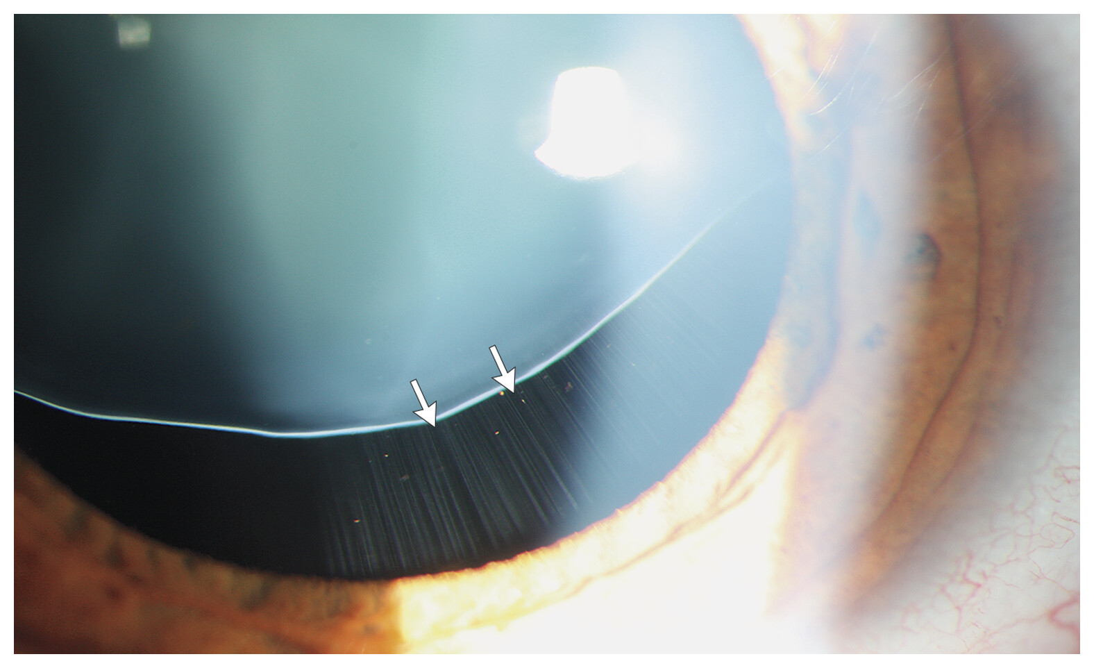
- The edge of the lens visible within the pupillary aperture, typically superotemporally
- High, often irregular astigmatism from the tilted lens
- Myopia, frequently significant, related to both axial elongation and the lens position
- Elevated risk of retinal detachment, related to the axial myopia and vitreous changes common in Marfan syndrome
All-fit Slit-Lamp Adapter
Record and share exactly what you see at the slit lamp. One adapter fits any slit lamp or surgical microscope — and any smartphone.
From Choroida — the team behind this siteSystemic Features That Should Prompt Consideration of Marfan Syndrome
- Tall stature with disproportionately long limbs and digits (arachnodactyly)
- Pectus deformity (excavatum or carinatum) and joint hypermobility
- Aortic root dilation and risk of dissection — the most life-threatening feature of the syndrome
- Mitral valve prolapse
- Dural ectasia and striae distensae (stretch marks) unrelated to weight change
Because aortic disease is the dominant driver of mortality in Marfan syndrome, an ophthalmologist who identifies characteristic ectopia lentis in a patient without a known diagnosis has a genuine responsibility to prompt cardiology referral and echocardiography, not just to manage the eye finding in isolation.
This is a genuine example of an ocular finding carrying life-or-death systemic significance well beyond the eye, since undiagnosed aortic root dilation can progress silently until a catastrophic dissection occurs, making the ophthalmologist’s referral potentially a life-saving intervention.
Differential Diagnosis of Ectopia Lentis
- Homocystinuria — lens typically subluxates inferonasally (opposite direction from Marfan), with additional risk of thromboembolism and developmental delay
- Weill-Marchesani syndrome — microspherophakia with lens subluxation, short stature (opposite body habitus from Marfan), and a higher risk of pupillary block glaucoma
- Traumatic lens subluxation — history of blunt trauma, usually unilateral and asymmetric
- Simple (familial) ectopia lentis — isolated zonular weakness without other systemic features
- Ehlers-Danlos syndrome — less consistently associated with ectopia lentis than the other conditions above
The direction of subluxation, body habitus, and associated systemic findings usually differentiate these causes without needing genetic testing, though molecular confirmation of FBN1 mutations supports the diagnosis when the clinical picture is ambiguous or when family screening is being planned, since a confirmed genetic diagnosis directly informs recurrence risk for future children.
Management
Mild subluxation with preserved vision through the native lens (aided by dilating the pupil optically, or working around the displaced optical zone with spectacle correction) can often be managed without surgery for extended periods, with regular monitoring for progression and for the retinal detachment risk that comes with the associated myopia.
Surgical lens removal is indicated when subluxation significantly affects vision, when the lens threatens to dislocate fully into the vitreous or anterior chamber, or when it contributes to secondary glaucoma, with the decision to operate generally weighed against the option of continued observation and optical management for as long as that remains functionally adequate.
Lensectomy in these eyes is technically more demanding than routine cataract surgery because the weak zonules cannot be relied upon for capsular support, and techniques often involve specialized capsular tension rings or segments, or intentional removal of the capsular bag with scleral- or iris-fixated intraocular lens placement.
Because the zonular weakness is a lifelong feature, these eyes need long-term follow-up for both the fellow eye and for late complications such as further lens or IOL decentration, since the same underlying connective tissue fragility that caused the original subluxation continues to affect any fixation device placed during surgery.


Document what you see
Two smartphone imaging tools built for everyday clinic use — one for the slit lamp, one for the fundus.
From Choroida — the team behind this siteReferences
- Loeys BL, Dietz HC, Braverman AC, et al. The revised Ghent nosology for the Marfan syndrome. Journal of Medical Genetics.
- Nemet AY, Assia EI, Apple DJ, Barequet IS. Current concepts of ocular manifestations in Marfan syndrome. Survey of Ophthalmology.
- Konradsen TR, Zetterström C. A descriptive study of ocular characteristics in Marfan syndrome. Acta Ophthalmologica.
- American Academy of Ophthalmology. Basic and Clinical Science Course, Section 11: Lens and Cataract.
Test yourself
A few questions straight from this article.
-
In which direction does the crystalline lens typically subluxate in Marfan syndrome?
The Marfan lens classically drifts up and out, and this direction is distinctive enough to help separate it from other causes of ectopia lentis. -
Which gene and protein underlie the zonular weakness of Marfan syndrome?
Fibrillin-1 is a major structural component of the zonular fibres, so FBN1 mutations leave the zonules fragmented and unable to hold the lens in place under normal mechanical stress. -
Why do patients with ectopia lentis in Marfan syndrome usually report a second image rather than sudden profound vision loss?
Because the displaced lens edge remains near the visual axis instead of falling back into the vitreous, patients describe monocular diplopia and gradual blur rather than an abrupt loss. -
Which pair of signs reflects loss of normal zonular support in ectopia lentis?
Tremulousness of the iris and of the lens on eye movement is the direct clinical expression of zonules that no longer anchor the lens firmly. -
Why are eyes with ectopia lentis in Marfan syndrome at elevated risk of retinal detachment?
The significant axial myopia of Marfan syndrome, combined with the vitreous changes that accompany it, raises detachment risk and is part of why these eyes need ongoing retinal surveillance. -
Which features point to homocystinuria rather than Marfan syndrome as the cause of ectopia lentis?
Homocystinuria displaces the lens in the opposite direction to Marfan syndrome and carries added risks of thromboembolism and developmental delay. -
Which combination characterises Weill-Marchesani syndrome as a cause of lens subluxation?
Weill-Marchesani combines a small spherical lens with the opposite body habitus to Marfan syndrome and a notably higher risk of pupillary block glaucoma. -
An ophthalmologist finds characteristic ectopia lentis in a patient with no systemic diagnosis. What is the essential next step?
Aortic root dilation is the dominant driver of mortality in Marfan syndrome and can progress silently to dissection, so prompting cardiac assessment may be life-saving. -
Which situation indicates surgical lens removal in ectopia lentis?
Surgery is reserved for subluxation that significantly impairs vision, lenses at risk of dislocating into the vitreous or anterior chamber, and eyes developing secondary glaucoma. -
Why is lensectomy in Marfan syndrome more demanding than routine cataract surgery?
Because the zonules cannot be trusted, surgeons use capsular tension rings or segments, or remove the bag and place a scleral- or iris-fixated intraocular lens.

Writes and reviews the clinical articles on Educate. Every post is checked against peer-reviewed sources before it goes live.


