
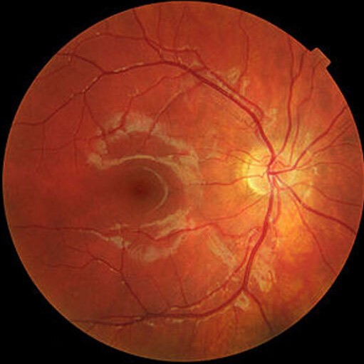
Photodynamic therapy (PDT) with verteporfin uses a photosensitizing drug and a nonthermal laser to close abnormal choroidal vessels while sparing most of the overlying retina.
It was the first effective treatment for neovascular AMD, and it lost that role to anti-VEGF injections.
It remains valuable in specific conditions, notably polypoidal choroidal vasculopathy, chronic central serous chorioretinopathy, and circumscribed choroidal hemangioma, where its effect on choroidal vessels cannot be matched by injections.
How Photodynamic Therapy Works
Verteporfin is a benzoporphyrin derivative given by intravenous infusion.
It circulates bound to lipoproteins and accumulates in vessels with high lipoprotein uptake, such as neovascular endothelium.
A nonthermal 689 nm laser then activates the drug, which generates singlet oxygen and other reactive species.
These damage endothelial cells and cause thrombosis and vascular occlusion in the treated area.
The standard protocol is 6 mg/m² of verteporfin infused over 10 minutes, followed by laser application at 15 minutes after the start of infusion, delivering 50 J/cm² over 83 seconds.
Reduced-dose and reduced-fluence protocols use half the drug dose or half the light energy, and they are used in chronic central serous chorioretinopathy to limit choroidal ischemia.
Historical Role in Neovascular AMD
The TAP study showed that verteporfin PDT reduced the risk of moderate visual loss in eyes with predominantly classic subfoveal choroidal neovascularization, compared with placebo.
Results in occult disease were less favorable.
Anti-VEGF drugs later produced actual visual gains, where PDT had only reduced the rate of loss, and PDT was largely replaced as monotherapy for neovascular AMD.
Fundus Explorer Pro
Photograph the retinal findings described here with the phone already in your pocket — 22 D optics and built-in illumination in one handheld unit.
From Choroida — the team behind this siteCurrent Indications
Polypoidal Choroidal Vasculopathy
PDT closes the polypoidal lesions, while anti-VEGF treats the branching vascular network and leakage (see polypoidal choroidal vasculopathy).
In the EVEREST II trial, ranibizumab combined with PDT produced greater visual gains and a higher rate of complete polyp regression than ranibizumab alone, and it required fewer injections.
Combination therapy or PDT alone is used depending on availability and the response.
Chronic Central Serous Chorioretinopathy
Half-dose or half-fluence PDT reduces choroidal hyperpermeability and resolves subretinal fluid.
In the PLACE trial, half-dose PDT led to complete resolution of subretinal fluid in about half of eyes, compared with about one-third after high-density subthreshold micropulse laser (see central serous chorioretinopathy).
A randomized trial of half-dose PDT for acute disease also reported faster fluid resolution than placebo.
Indications include persistent fluid beyond three to four months, recurrent disease, and macular damage in a symptomatic eye.
Circumscribed Choroidal Hemangioma
PDT closes the vascular tumor, causes resolution of subretinal fluid, and often improves vision (see choroidal hemangioma).
It has become the first-line treatment for symptomatic circumscribed choroidal hemangioma.
Other Uses
- Myopic and other secondary choroidal neovascularization in selected patients (see myopic CNV)
- Pachychoroid neovasculopathy, usually with anti-VEGF (see pachychoroid neovasculopathy)
- Retinal vasoproliferative tumors and some choroidal osteomas with neovascularization
Patient Selection and Preparation
- Confirm the diagnosis and the target lesion with OCT, fluorescein angiography, and indocyanine green angiography
- Check for allergies to verteporfin, and for hepatic disease, porphyria, and pregnancy
- Check the patient’s ability to avoid bright light for 48 hours after infusion
- Measure body height and weight to calculate the body surface area
- Obtain consent for the small risk of vision loss
Angiography guides the treatment spot, which should include the polyps or the area of leakage and a margin.
Adverse Effects
- Photosensitivity: patients must avoid direct sunlight and bright indoor light for 48 hours, wearing protective clothing and dark glasses
- Infusion reactions: back pain, chest pain, and infusion-site extravasation
- Acute severe visual loss: a small percentage of patients, usually within a week
- Choroidal ischemia and RPE atrophy: particularly with full-fluence protocols
- Subretinal, retinal, or vitreous hemorrhage: especially in polypoidal disease
- Secondary choroidal neovascularization after treatment of chronic central serous chorioretinopathy
Reduced-fluence protocols reduce these risks, and the treatment area should be limited to the target lesion.
Combination Strategies
PDT is often combined with anti-VEGF injections in eyes that have both vascular closure and leakage to treat.
- In polypoidal disease, the injection is usually given on the same day or a few days before PDT, and the response is judged by ICGA and OCT
- In pachychoroid neovasculopathy, the anti-VEGF component treats the neovascular network and PDT reduces the choroidal hyperpermeability that drives it
- In chronic central serous chorioretinopathy with secondary neovascularization, combination therapy is used when fluid persists after PDT
The order of treatment and the number of sessions are individualized, since evidence for optimal sequencing is limited.
Limitations
PDT does not treat the intraretinal and subretinal fluid produced by VEGF alone, and it does not restore lost photoreceptors.
It works through selective vascular closure and may need repeat sessions.
Availability of verteporfin has been limited in some regions, which restricts its use.
Anti-VEGF therapy is preferred for most types of neovascular macular degeneration, and PDT is used in combination for selected lesions.
Prognosis
In appropriate patients, PDT gives durable resolution of fluid and stabilizes or improves vision.
Outcomes are best in eyes without established RPE atrophy, and treatment early in the disease preserves the most vision.


Document what you see
Two smartphone imaging tools built for everyday clinic use — one for the slit lamp, one for the fundus.
From Choroida — the team behind this siteReferences
- Treatment of Age-Related Macular Degeneration With Photodynamic Therapy (TAP) Study Group. Photodynamic therapy of subfoveal choroidal neovascularization in age-related macular degeneration with verteporfin: one-year results of 2 randomized clinical trials. TAP report 1. Arch Ophthalmol. 1999;117:1329-1345.
- Koh A, Lai TYY, Takahashi K, et al. Efficacy and safety of ranibizumab with or without verteporfin photodynamic therapy for polypoidal choroidal vasculopathy: a randomized clinical trial (EVEREST II). JAMA Ophthalmol. 2017;135:1206-1213.
- van Dijk EHC, Fauser S, Breukink MB, et al. Half-dose photodynamic therapy versus high-density subthreshold micropulse laser treatment in patients with chronic central serous chorioretinopathy: the PLACE trial. Ophthalmology. 2018;125:1547-1555.
- Chan WM, Lai TYY, Lai RYK, Tang EWH, Liu DTL, Lam DSC. Half-dose verteporfin photodynamic therapy for acute central serous chorioretinopathy: one-year results of a randomized controlled trial. Ophthalmology. 2008;115:1756-1765.
- Schmidt-Erfurth U, Hasan T. Mechanisms of action of photodynamic therapy with verteporfin for the treatment of age-related macular degeneration. Surv Ophthalmol. 2000;45:195-214.
Test yourself
A few questions straight from this article.
-
How does photodynamic therapy with verteporfin close abnormal choroidal vessels?
A nonthermal 689 nm laser activates verteporfin, generating singlet oxygen and other reactive species that damage endothelial cells and cause thrombosis and vascular occlusion in the treated area. -
Why does intravenous verteporfin concentrate in neovascular tissue?
Verteporfin is a benzoporphyrin derivative that circulates bound to lipoproteins and accumulates in vessels with high lipoprotein uptake, such as neovascular endothelium. -
What is the standard full-dose verteporfin regimen for photodynamic therapy?
The standard protocol is 6 mg/m² of verteporfin infused over 10 minutes, with laser applied 15 minutes after the start of infusion delivering 50 J/cm² over 83 seconds. -
Why are reduced-dose or reduced-fluence protocols preferred in chronic central serous chorioretinopathy?
Reduced-dose and reduced-fluence protocols use half the drug dose or half the light energy and are used in chronic central serous chorioretinopathy to limit choroidal ischaemia. -
What did the TAP study demonstrate for verteporfin photodynamic therapy in neovascular AMD?
TAP showed verteporfin photodynamic therapy reduced the risk of moderate visual loss in predominantly classic subfoveal choroidal neovascularisation compared with placebo, with less favourable results in occult disease. -
What did EVEREST II show about ranibizumab combined with photodynamic therapy in polypoidal choroidal vasculopathy?
In EVEREST II, ranibizumab combined with photodynamic therapy produced greater visual gains and a higher rate of complete polyp regression than ranibizumab alone, and required fewer injections. -
In the PLACE trial, how did half-dose photodynamic therapy compare with subthreshold micropulse laser?
In PLACE, half-dose photodynamic therapy gave complete resolution of subretinal fluid in about half of eyes, compared with about one-third after high-density subthreshold micropulse laser. -
What is the first-line treatment for symptomatic circumscribed choroidal haemangioma?
Photodynamic therapy closes the vascular tumour, resolves subretinal fluid and often improves vision, and it has become first-line treatment for symptomatic circumscribed choroidal haemangioma. -
What precaution must a patient follow after a verteporfin infusion?
Verteporfin causes photosensitivity, so patients must avoid direct sunlight and bright indoor light for 48 hours, wearing protective clothing and dark glasses. -
Which limitation explains why anti-VEGF therapy is preferred for most neovascular macular degeneration?
Photodynamic therapy works by selective vascular closure, so it does not clear the intraretinal and subretinal fluid produced by VEGF alone and does not restore lost photoreceptors.

Writes and reviews the clinical articles on Educate. Every post is checked against peer-reviewed sources before it goes live.


