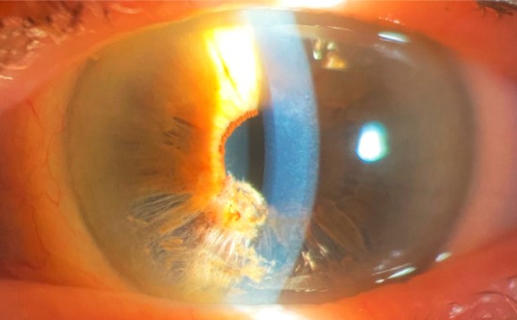
Iridoschisis is a rare degenerative condition in which the anterior stromal layer of the iris splits from the posterior layer, producing visible strands or sheets of anterior stroma that separate and float freely within the anterior chamber.
It is almost always seen in elderly patients and is frequently, though not universally, associated with angle-closure glaucoma, which is usually what actually brings the finding to clinical attention rather than the iris change itself.
Distinguishing iridoschisis from other causes of iris strands and atrophy, most notably essential iris atrophy within the ICE syndrome spectrum, matters because the two conditions differ in age of onset, laterality, and overall clinical context.
Iridoschisis is genuinely rare, and most ophthalmologists will encounter it only occasionally over a career, which makes it easy to misclassify on first exposure without a specific frame of reference for the finding.
Clinical Features
- Splitting of the anterior iris stroma from the posterior layers, producing visible fibrillar strands that can wave freely in the anterior chamber with eye movement
- Typical onset in elderly patients, usually in the seventh decade of life or later, distinguishing it from the younger age of onset typical of ICE syndrome
- Frequent bilateral involvement, though often asymmetric in severity between the two eyes
- Strong association with angle-closure glaucoma, present in a substantial proportion of affected patients at the time of diagnosis
Pathophysiology
The exact mechanism remains incompletely understood, but iridoschisis is generally considered a degenerative, age-related process affecting the structural integrity of the iris stroma rather than an inflammatory or infectious one.
The strong association with angle closure has led to the theory that chronic or intermittent elevated pressure, or the mechanical stress from a crowded, narrow anterior chamber, contributes to splitting an already age-weakened iris stroma, though whether angle closure is a cause or simply a frequent companion condition remains debated.
Some cases occur without any associated angle-closure component, suggesting that intrinsic age-related stromal degeneration alone may be sufficient to produce the finding in at least a subset of patients.
Histopathologic studies have shown thinning and fragmentation of the anterior stromal layer with relative preservation of the posterior pigmented epithelium, consistent with a primary stromal rather than epithelial process.
All-fit Slit-Lamp Adapter
Record and share exactly what you see at the slit lamp. One adapter fits any slit lamp or surgical microscope — and any smartphone.
From Choroida — the team behind this siteDifferential Diagnosis
Essential iris atrophy, part of the iridocorneal endothelial syndrome spectrum discussed in its own dedicated article on this site, typically affects younger to middle-aged patients, is characteristically unilateral, and arises from a primary corneal endothelial abnormality rather than isolated iris stromal splitting.
Iris strands from prior trauma or surgery have an identifiable inciting history and a different, more localized pattern than the diffuse, bilateral changes typical of age-related iridoschisis.
Pseudoexfoliation syndrome, discussed in its own dedicated article on this site, can coexist with angle-closure glaucoma in an elderly patient but produces a different pattern of findings, with characteristic flaky material on the lens and pupillary margin rather than splitting of the iris stroma itself.
Clinical Significance
The visible iris strands themselves are not typically the primary threat to vision; rather, the associated angle-closure glaucoma, when present, is what actually drives the urgency of management and the risk to the patient’s sight.
Free-floating iris strands can occasionally interfere mechanically with angle structures or contribute to intermittent pupillary block, adding a structural component to the glaucoma risk beyond the general anatomic crowding common in elderly angle-closure-prone eyes.
Recognizing iridoschisis should prompt a careful gonioscopic assessment of the angle, given how frequently the two findings occur together and how much the glaucoma management, rather than the iris finding itself, actually matters for the patient’s long-term visual outcome.
Management
There is no specific treatment directed at the iris splitting itself, since the strands are not typically symptomatic or visually significant on their own.
Management is centered entirely on identifying and treating any associated angle-closure glaucoma, following standard approaches including laser peripheral iridotomy or, when indicated, other glaucoma-specific interventions discussed in their own dedicated articles on this site.
Regular monitoring for glaucoma, given the strong association between the two conditions, is warranted in any patient diagnosed with iridoschisis, even when angle closure is not present at the time of initial diagnosis.
Patients can understandably find the visible, mobile iris strands alarming to notice themselves, and simple reassurance that the strands alone are not a direct threat to vision, distinct from the glaucoma risk that actually needs monitoring, is a useful part of counseling at diagnosis.


Document what you see
Two smartphone imaging tools built for everyday clinic use — one for the slit lamp, one for the fundus.
From Choroida — the team behind this siteReferences
- Rodrigues MM, Streeten BW, Spaeth GL, et al. Iridoschisis: histopathology, ultrastructure, and clinical review. Ophthalmology.
- American Academy of Ophthalmology. Basic and Clinical Science Course, Section 10: Glaucoma.
- Bowling B. Kanski’s Clinical Ophthalmology.
Test yourself
A few questions straight from this article.
-
Iridoschisis is defined by separation between which iris layers?
The anterior stromal layer splits away from the posterior layers, leaving strands or sheets of stroma that float freely in the anterior chamber. -
At what stage of life does iridoschisis typically become apparent?
Onset is typically in elderly patients, usually the seventh decade or later, which is one of the features separating it from ICE syndrome. -
What is the usual laterality of iridoschisis?
Involvement is frequently bilateral, although the severity commonly differs between the two eyes. -
Which type of glaucoma is strongly associated with iridoschisis?
Angle-closure glaucoma accompanies iridoschisis in a substantial proportion of patients, and it is usually the glaucoma rather than the iris change that brings them in. -
Iridoschisis is best understood as which kind of disease process?
It is regarded as an age-related degenerative loss of iris stromal structural integrity rather than an inflammatory or infectious disease. -
Histopathology of iridoschisis shows which pattern of iris change?
Thinning and fragmentation affect the anterior stromal layer while the posterior pigmented epithelium is relatively preserved, confirming a primary stromal process. -
Which feature favours essential iris atrophy over iridoschisis?
Essential iris atrophy within the ICE spectrum is characteristically unilateral, affects younger to middle-aged patients, and stems from a primary corneal endothelial abnormality. -
Beyond anatomic crowding, free-floating iris strands in iridoschisis may contribute to what?
The mobile strands can mechanically interfere with angle structures or contribute to intermittent pupillary block, adding a structural element to the glaucoma risk. -
A patient with iridoschisis whose angles are open at diagnosis requires what follow-up?
Because the association with angle closure is strong, glaucoma surveillance is warranted even when the angle is not closed at the time of initial diagnosis. -
Management of iridoschisis is centred on which goal?
No treatment is directed at the splitting itself; care focuses on identifying and treating angle-closure glaucoma, with laser peripheral iridotomy where indicated.

Writes and reviews the clinical articles on Educate. Every post is checked against peer-reviewed sources before it goes live.