
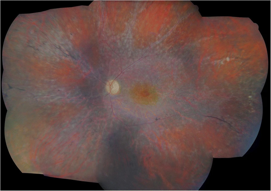
Cancer-associated retinopathy is a paraneoplastic autoimmune condition in which antibodies generated against a tumor cross-react with retinal proteins, causing progressive, often rapid and bilateral, vision loss that can precede the diagnosis of the underlying malignancy itself, making ophthalmic recognition of this pattern genuinely important for early cancer detection in some patients.
The Paraneoplastic Mechanism
Cancer-associated retinopathy arises when a tumor expresses a protein that is also normally expressed in retinal cells, most classically recoverin, a calcium-binding protein involved in phototransduction.
The immune system, generating antibodies against the tumor-expressed protein as part of an anti-tumor immune response, produces antibodies that cross-react with the same protein in the retina, attacking retinal photoreceptors as an unintended consequence of what began as an immune response against the cancer, in a molecular mimicry mechanism.
Associated Malignancies
Small cell lung cancer is the malignancy most classically associated with cancer-associated retinopathy, particularly in cases involving anti-recoverin antibodies, though the syndrome has also been described with gynecologic, breast, and other malignancies, and a range of other retinal and bipolar cell antigens beyond recoverin have been implicated in some cases.
Fundus Explorer Pro
Photograph the retinal findings described here with the phone already in your pocket — 22 D optics and built-in illumination in one handheld unit.
From Choroida — the team behind this siteClinical Features
- Progressive, often rapidly evolving, bilateral vision loss, sometimes developing over weeks to a few months
- Photopsias (flickering or shimmering light sensations), often an early symptom
- Difficulty with dark adaptation and night vision, reflecting the rod photoreceptor involvement typical of anti-recoverin disease
- Visual field constriction, sometimes progressing to severe peripheral field loss
- The fundus examination can appear relatively normal, particularly early in the disease course, which is part of why the degree of functional visual loss often seems disproportionate to the visible retinal findings on routine examination
- Retinal vessel attenuation and pigmentary changes can develop as the disease progresses
Diagnostic Evaluation
- Electroretinography is central to diagnosis, typically showing markedly reduced or extinguished responses, often affecting both rod and cone function, out of proportion to the funduscopic appearance, a finding that strongly supports a diffuse photoreceptor dysfunction process such as cancer-associated retinopathy
- Serum testing for anti-retinal antibodies, including anti-recoverin, though interpretation requires care given both false positives and the existence of cancer-associated retinopathy cases without detectable anti-recoverin antibodies
- A thorough search for an underlying malignancy when cancer-associated retinopathy is suspected, including age-appropriate cancer screening and imaging directed by clinical suspicion, particularly for small cell lung cancer given its strong association
- In a patient with a known cancer history, recognizing new, otherwise unexplained progressive visual loss as a possible paraneoplastic phenomenon, rather than assuming an unrelated ophthalmic cause
Differential Diagnosis
Cancer-associated retinopathy must be distinguished from other causes of progressive retinal dysfunction, including inherited retinal degenerations such as retinitis pigmentosa, other autoimmune retinopathies not associated with malignancy, and toxic or nutritional causes of diffuse photoreceptor dysfunction, with the pace of onset, bilaterality, and electroretinogram pattern helping guide this distinction.
Management
Treating the Underlying Malignancy
Identifying and treating the underlying cancer is a central part of management, both for the patient’s overall prognosis and because tumor treatment can, in some cases, be associated with stabilization of the retinopathy, though visual recovery is often limited even with successful cancer treatment given the degree of photoreceptor damage that may have already occurred.
Immunomodulatory Treatment
Systemic corticosteroids, intravenous immunoglobulin, and other immunosuppressive approaches have been used in an attempt to slow or halt progression of the retinopathy, with variable and generally modest reported success, reflecting the aggressive nature of this autoimmune process once established.
Prognosis
Cancer-associated retinopathy tends to follow an aggressive course, and visual loss is often severe and, once established, largely irreversible, even with treatment of the underlying cancer and attempts at immunomodulation.
Early recognition, particularly in a patient not yet known to have cancer, carries importance beyond the eye itself, since it can prompt a cancer workup that identifies a treatable malignancy at an earlier, more favorable stage than it might otherwise have been found.


Document what you see
Two smartphone imaging tools built for everyday clinic use — one for the slit lamp, one for the fundus.
From Choroida — the team behind this siteReferences
- Thirkill CE, FitzGerald P, Sergott RC, Roth AM, Tyler NK, Keltner JL. Cancer-associated retinopathy (CAR syndrome) with antibodies reacting with retinal, optic-nerve, and cancer cells. N Engl J Med. 1989;321:1589-1594.
- Adamus G. Autoantibody targets and their cancer relationship in the pathogenicity of paraneoplastic retinopathy. Autoimmun Rev. 2009;8:410-414.
- Grewal DS, Fishman GA, Jampol LM. Autoimmune retinopathy and antiretinal antibodies: a review. Retina. 2014;34:827-845.
- Chan JW. Paraneoplastic retinopathies and optic neuropathies. Surv Ophthalmol. 2003;48:12-38.
Test yourself
A few questions straight from this article.
-
What mechanism damages the retina in cancer-associated retinopathy?
A tumour expresses a protein also found in retinal cells, and the antibodies raised against it attack the photoreceptors by molecular mimicry. -
What is recoverin, the classic antigen in cancer-associated retinopathy?
Recoverin is a calcium-binding protein of the phototransduction cascade, expressed both in retinal cells and by the tumour that triggers the antibody response. -
Which malignancy is most classically associated with cancer-associated retinopathy?
Small cell lung cancer is the classic association, particularly in cases with anti-recoverin antibodies, although gynaecologic, breast and other cancers are also described. -
Which symptom is often an early feature of cancer-associated retinopathy?
Photopsias are often an early symptom, alongside progressive bilateral visual loss developing over weeks to a few months. -
Why do patients with anti-recoverin retinopathy report poor night vision?
Difficulty with dark adaptation and night vision reflects the rod photoreceptor involvement typical of anti-recoverin disease. -
Which test is central to diagnosing cancer-associated retinopathy?
The electroretinogram is typically markedly reduced or extinguished in both rod and cone function, out of proportion to a fundus that can look relatively normal. -
Why must anti-retinal antibody results be interpreted with care in suspected cancer-associated retinopathy?
Serum anti-retinal antibody testing is useful but imperfect: false positives occur, and cancer-associated retinopathy exists without detectable anti-recoverin antibodies. -
Which features help separate cancer-associated retinopathy from retinitis pigmentosa?
The rapid pace of onset, bilateral involvement and the electroretinogram pattern guide the distinction from inherited degenerations and from toxic or nutritional causes. -
What effect can treating the underlying malignancy have on cancer-associated retinopathy?
Tumour treatment can in some cases be associated with stabilisation of the retinopathy, but visual recovery is often limited because photoreceptor damage has already occurred. -
Why does early recognition of cancer-associated retinopathy matter beyond the eye?
Because the retinopathy can precede the cancer diagnosis, recognising it may lead to a workup that finds a treatable malignancy at a more favourable stage.

Writes and reviews the clinical articles on Educate. Every post is checked against peer-reviewed sources before it goes live.


