
CASE REPORT
A 36-year-old female presented with complaints of gradually progressive defective vision for 3 years, which was more in the left eye (OS) than the right eye (OD). She had no history of using glasses in the past.

On examination, the patient had uncorrected visual acuity of 3/60 OD and 2/60 OS, which improved with 19 DS bilaterally to 6/60 in OD and to 5/60 in OS. Intraocular pressures were measured as 18 mmHg in both eyes (OU).
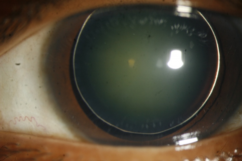
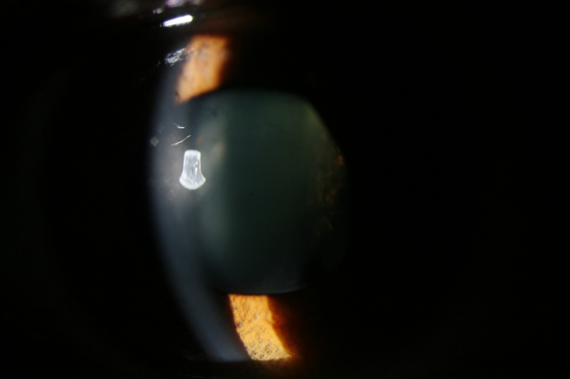
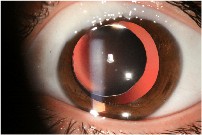
Slit-lamp examination revealed iridodonesis OU. On complete pupillary dilation, OU revealed a spherical cataractous lens of nucleus sclerosis Grade 3, associated with gross phacodonesis. The edges of the lens were visible, with thin stretched-out zonules OU, however, there was no gross subluxation a and b.
Gonioscopic examination OU showed highly pigmented angles with minimal peripheral anterior synechiae. Posterior segment examination was within normal limits OU. During optical biometry, lens thickness was found to be about 4.58 mm OD and 5.09 mm OS.
The anterior chamber depths were about 3.08 mm OD and 3.02 mm OS, with axial lengths of 22.27 mm and 21.99 mm, respectively, in her OD and OS. Her systemic workup, including serology, was found to be within normal limits.
Her entire family was screened for the same and was found to have no similar ocular findings. The patient was diagnosed with bilateral isolated Microspherophakia (MSP).
Microspherophakia: DISEASE entity
Microspherophakia (MSP) is characterized by increased anteroposterior diameter and reduced equatorial diameter of the crystalline lens. It is a rare condition, usually bilateral.
Microspherophakia has the following characteristics:
- It is usually bilateral
- The small diameter of the lens
- Increased anteroposterior diameter of the lens
- The Equator of the lens is visible with full mydriasis
- Movement of the lens with a change in posture
- May be associated with lens dislocation/ subluxation, high myopia, defective accommodation, glaucoma
Genetics:
Mutations in the following genes have been noted:
- Latent TGF β-binding protein-2 (LTBP2) gene
- ADAMTS17
LTBPs are a family of proteins showing structural homologies with fibrillins and are expressed in the trabecular meshwork, ciliary processes, and lens capsule/epithelium layer. It might have a role in the regulation of elastic fiber assembly.
ADAMTS17 is part of the same family of metalloproteinases as ADAMTS10 which is the main gene associated with Weill-Marchesani syndrome.
Microspherophakia MANAGEMENT
Medical and surgical management of complications seen with microspherophakia may be difficult.
Cycloplegics are the treatment of choice. Miotics if given in microspherophakia might lead to pupillary block glaucoma.
Stepwise protocol for the treatment of glaucoma in microspherophakia:
- Nd: Yag Laser Peripheral iridotomy (although this may have limited effect)
- Clear lens extraction with or without goniosynechiolysis
- Filtering surgery
- Tube shunt surgery
Indications of clear lens extraction in patients with microspherophakia:
- Corneo-lenticular contact
- Unilateral high myopia
- Pupillary block
- Secondary intractable glaucoma
Intraoperative complications associated with clear lens extraction in these patients include difficulty in performing capsulorhexis and implanting IOL due to smaller bag sizes.
HOW TO TAKE SLIT-LAMP EXAM IMAGES WITH A SMARTPHONE?
Smartphone slit-lamp photography is the new advancement in the field of science and technology in which photographs of the desired slit-lamp finding can be taken with smartphones by using the slit-lamp adapters.
Slit-lamp Smartphone photography
REFERENCES
- Şimşek T, Beyazyıldız E, Şimşek E, Öztürk F. Isolated Microspherophakia Presenting with Angle-Closure Glaucoma. Türk Oftalmoloji Dergisi. 2016;46(5):237-240
- Bitar MS, Farooq AV, Abbasian J (2016) Challenges in Diagnosing Microspherophakia in a Pediatric Patient. JSM Ophthalmol 4(1): 1040
- Senthil S, Rao HL, Hoang NT, et al. Glaucoma in microspherophakia: presenting features and treatment outcomes. J Glaucoma. 2014. Apr-May;23(4):262-7.
- Muralidhar R, Ankush K, Vijayalakshmi P, George V. Visual outcome and incidence of glaucoma in patients with microspherophakia. Eye. 2014;29(3):350-355
- Medhi J, DasGupta S, Bhattacharjee H, Bhattacharjee K. Clear lens extraction and intraocular lens implantation in a case of microspherophakia with secondary angle closure glaucoma. Indian Journal of Ophthalmology. 2010;58(1):67.
