The name describes what is missing, but the disease is really about everything else that goes wrong when that one structure never forms properly.
Aniridia is congenital absence or near-absence of the iris, and it is far more than a cosmetic finding.
The same gene mutation responsible for the missing iris also disrupts development of the cornea, lens, and fovea.
Vision loss in aniridia usually has little to do with the absent iris itself and everything to do with these associated structural problems.
A subset of cases signals an underlying chromosomal deletion with serious systemic implications, including a real risk of kidney cancer in early childhood.
Recognizing aniridia and its associated syndrome is what triggers the systemic workup that can be genuinely life-saving.
What Is Aniridia?
Aniridia is a panocular developmental disorder characterized by complete or partial absence of the iris, resulting from mutation of the PAX6 gene.
Its defining features include:
- A variable degree of iris absence, from a thin rudimentary stump to complete absence
- Bilateral involvement in almost all cases
- Associated developmental abnormalities of the cornea, lens, and fovea, since PAX6 governs eye development broadly, not just the iris
- Two main forms — isolated (familial) aniridia and aniridia associated with WAGR syndrome
Because PAX6 is a master regulatory gene for eye development, aniridia is best understood as a whole-eye developmental disorder that happens to be named after its most visible feature.
Epidemiology
Aniridia is a rare condition, with a well-characterized genetic basis.
- Roughly two-thirds of cases are familial, inherited in an autosomal dominant pattern with high penetrance
- The remaining cases are sporadic, and a subset of sporadic cases result from a contiguous gene deletion at chromosome 11p13 that also removes the adjacent WT1 gene
- This 11p13 deletion produces WAGR syndrome — Wilms tumor, Aniridia, Genitourinary anomalies, and Range of developmental delay
- Any child with sporadic (non-familial) aniridia requires genetic testing to determine WAGR risk
Distinguishing familial from sporadic aniridia is one of the most consequential distinctions in pediatric ophthalmology, given the Wilms tumor risk tied to the sporadic, deletion-associated form.
All-fit Slit-Lamp Adapter
Record and share exactly what you see at the slit lamp. One adapter fits any slit lamp or surgical microscope — and any smartphone.
From Choroida — the team behind this sitePathophysiology
PAX6 functions as a master control gene during early eye development.
- PAX6 mutation or deletion disrupts normal development of ocular structures derived from neuroectoderm and surface ectoderm
- Iris development is arrested early, leaving a rudimentary stump or no iris tissue at all
- The same disruption impairs normal corneal limbal stem cell development, predisposing to progressive aniridia-associated keratopathy later in life
- Foveal development is frequently incomplete (foveal hypoplasia), which is often the true limiting factor for visual acuity
- Associated lens abnormalities (cataract, ectopia lentis) and glaucoma commonly develop as secondary complications over time
Because a single gene governs development of so many ocular structures simultaneously, aniridia is rarely just about the iris — it is a syndrome of the whole developing eye.
Risk Factors
Genetic Risk Factors
- A parent with familial aniridia, given autosomal dominant inheritance with high penetrance
- De novo PAX6 mutation or 11p13 deletion in sporadic cases
Associated Systemic Risk
- Sporadic aniridia carries meaningful risk of an underlying 11p13 deletion and WAGR syndrome, including Wilms tumor
- Any child with sporadic, non-familial aniridia needs prompt genetic testing and, if the deletion is confirmed, a structured Wilms tumor screening protocol
The urgency of ruling out WAGR syndrome in sporadic cases is what separates aniridia from most other congenital eye findings — this is one of the relatively few ophthalmic diagnoses that can directly trigger a childhood cancer screening protocol.
Clinical Presentation
Symptoms
- Poor vision from birth, often with nystagmus, related primarily to foveal hypoplasia rather than the absent iris
- Photophobia and glare from the absent iris diaphragm, which normally regulates light entry
- Progressive vision loss later in life if glaucoma or aniridia-associated keratopathy develops
Examination Findings
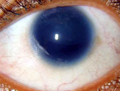
Aniridia: a uniformly large, dark pupillary aperture with complete absence of visible iris tissue, sclera bordering directly onto the corneal limbus- A large, uniformly dark pupillary aperture with absent or minimal visible iris tissue, sclera visible directly at the limbus
- Nystagmus, often present from infancy, related to foveal hypoplasia
- Corneal changes ranging from normal in early childhood to progressive limbal stem cell deficiency and corneal opacification later in life
- Cataract, lens subluxation, or glaucoma may be present, particularly with increasing age
The absence of a normal iris diaphragm is immediately obvious on external exam, but the associated foveal hypoplasia — usually not directly visible without dilated exam or OCT — is what actually drives most of the vision limitation.
Diagnostic Evaluation
Ocular Examination
- Dilated fundus examination and OCT to assess for foveal hypoplasia and optic nerve involvement
- Regular intraocular pressure monitoring given the significant lifetime glaucoma risk
- Serial corneal surface examination to monitor for aniridia-associated keratopathy
Genetic Testing
- PAX6 mutation analysis and testing for the 11p13 deletion, particularly in sporadic (non-familial) cases
- Genetic counseling for the family once the underlying mutation is characterized
Systemic Screening (When Indicated)
- Renal ultrasound screening for Wilms tumor in any child with a confirmed 11p13 deletion, following a structured surveillance schedule through early childhood
- Assessment for genitourinary anomalies and developmental delay as part of the broader WAGR evaluation
Genetic testing is not optional in sporadic aniridia — it is what determines whether a child needs a childhood cancer surveillance program.
Differential Diagnosis
Conditions that can resemble aniridia include:
- Traumatic iris loss or extensive iridodialysis — an acquired history of trauma rather than a congenital, bilateral presentation
- Axenfeld-Rieger syndrome — iris abnormalities and corectopia, but typically with residual iris tissue and characteristic angle anomalies rather than complete absence
- Large surgical iridectomy or aggressive iris coloboma — a partial, sector-shaped defect rather than the near-total absence seen in aniridia
The bilateral, congenital, near-total absence of iris tissue is distinctive enough that aniridia is rarely confused with these mimics on careful examination.
Management
Optical and Low Vision Management
- Tinted contact lenses or glasses with an artificial iris/pupil aperture to reduce photophobia and glare
- Low vision aids tailored to the degree of visual impairment from foveal hypoplasia
Monitoring and Managing Complications
- Regular glaucoma screening and treatment, since elevated IOP is a common and significant complication over a patient’s lifetime
- Monitoring and staged management of cataract when present, often surgically more complex given the absent iris support
- Ocular surface management for aniridia-associated keratopathy, ranging from lubrication to limbal stem cell transplantation in advanced cases
Systemic Care Coordination
- Coordinated care with genetics, oncology (for WAGR-associated cases), and pediatrics
- Structured, scheduled renal ultrasound surveillance in children with a confirmed 11p13 deletion until the period of Wilms tumor risk has passed
Lifelong ophthalmic surveillance matters as much as any single treatment — most of the vision loss in aniridia accumulates gradually from glaucoma and keratopathy over decades, not from the absent iris itself.
Prognosis
Visual prognosis is highly variable and depends on the severity of associated ocular findings.
- Baseline vision is often limited by foveal hypoplasia and nystagmus, and does not typically improve with age
- Glaucoma and aniridia-associated keratopathy are the major drivers of progressive vision loss over a patient’s lifetime
- With WAGR syndrome, overall prognosis also depends on Wilms tumor screening and systemic outcomes, not just the eye findings
The eye exam alone never tells the whole story in aniridia — long-term outcome depends as much on genetic counseling and, where relevant, cancer surveillance as it does on anything done in the eye clinic.


Document what you see
Two smartphone imaging tools built for everyday clinic use — one for the slit lamp, one for the fundus.
From Choroida — the team behind this siteReferences
- Lee H, Khan R, O’Keefe M. Aniridia: current pathology and management. Acta Ophthalmologica. 2008.
- Hingorani M, Hanson I, van Heyningen V. Aniridia. European Journal of Human Genetics. 2012.
- Muto R, Yamamori S, Ohashi H, Osawa M. Prediction by FISH analysis of the occurrence of Wilms tumor in aniridia patients. American Journal of Medical Genetics. 2002.
- Aniridia. StatPearls, NCBI Bookshelf.
- Aniridia. EyeWiki, American Academy of Ophthalmology.
Test yourself
A few questions straight from this article.
-
Mutation of which gene causes aniridia?
Aniridia results from mutation of PAX6, a master regulatory gene for eye development, which is why the cornea, lens and fovea are affected as well as the iris. -
What is the typical laterality of aniridia?
Aniridia is bilateral in almost all cases, consistent with its origin in a developmental gene mutation rather than any localised or acquired insult. -
Approximately what proportion of aniridia cases are familial and autosomal dominant?
Roughly two-thirds of cases are familial, inherited in an autosomal dominant pattern with high penetrance; the remainder are sporadic and carry the risk of an underlying deletion. -
In aniridia, deletion of which adjacent gene at chromosome 11p13 produces WAGR syndrome?
A contiguous gene deletion at 11p13 removes PAX6 along with the adjacent WT1 gene, producing WAGR syndrome: Wilms tumor, aniridia, genitourinary anomalies and a range of developmental delay. -
Which associated abnormality is usually the true limiting factor for visual acuity in aniridia?
Foveal development is frequently incomplete in aniridia, and this foveal hypoplasia, together with the resulting nystagmus, is what mainly limits acuity rather than the missing iris. -
Which symptom in aniridia results directly from loss of the iris diaphragm?
The iris normally regulates how much light enters the eye, so its absence produces photophobia and glare, managed with tinted lenses or an artificial iris aperture. -
What mechanism underlies aniridia-associated keratopathy?
PAX6 disruption impairs normal corneal limbal stem cell development, so the ocular surface deteriorates over time into progressive limbal stem cell deficiency and corneal opacification. -
Which surveillance is required in a child with aniridia and a confirmed 11p13 deletion?
A confirmed 11p13 deletion triggers structured renal ultrasound surveillance for Wilms tumor through early childhood, which is why genetic testing in sporadic aniridia is not optional. -
Which condition is distinguished from aniridia by residual iris tissue and characteristic angle anomalies?
Axenfeld-Rieger syndrome produces iris abnormalities and corectopia but typically leaves residual iris tissue with characteristic angle anomalies, rather than the near-total absence seen in aniridia. -
Which two complications drive progressive vision loss over a lifetime in aniridia?
Baseline acuity is set by foveal hypoplasia and does not usually improve, so later decline comes from glaucoma and aniridia-associated keratopathy accumulating over decades.

Writes and reviews the clinical articles on Educate. Every post is checked against peer-reviewed sources before it goes live.