
Case Study
A 62-year-old woman presented with progressive, painless vision loss in both eyes over several months. She reported shimmering photopsias and night vision difficulties.
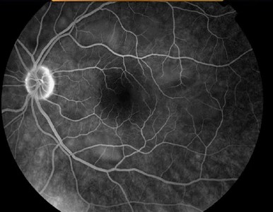
Her medical history included small-cell lung carcinoma diagnosed one year earlier, currently in remission. On examination, visual acuity was 20/80 in the right eye and 20/100 in the left eye.
Fundus examination showed mild retinal vascular attenuation with subtle retinal pigment epithelium (RPE) mottling. Optical coherence tomography (OCT) revealed outer retinal thinning with disruption of the ellipsoid zone.
Full-field electroretinography (ERG) demonstrated markedly reduced rod and cone responses. A diagnosis of paraneoplastic retinopathy was made, confirmed by detection of anti-recoverin antibodies in serum.
Immunosuppressive therapy with corticosteroids and intravenous immunoglobulin (IVIG) was initiated, though vision remained stable without significant improvement.
Disease Entity
Paraneoplastic retinopathy (PR) is a rare, immune-mediated retinal degeneration that occurs in association with systemic malignancies.
It results from cross-reactivity between tumor antigens and retinal proteins, leading to autoimmune destruction of photoreceptors and retinal cells.
PR represents a broader group of paraneoplastic syndromes, with cancer-associated retinopathy (CAR) and melanoma-associated retinopathy (MAR) as the two most well-recognized subtypes.
Fundus Explorer Pro
Photograph the retinal findings described here with the phone already in your pocket — 22 D optics and built-in illumination in one handheld unit.
From Choroida — the team behind this sitePathophysiology
The pathogenesis of PR is based on molecular mimicry. Tumors express antigens that resemble retinal proteins.
This antigenic similarity triggers the production of autoantibodies and autoreactive T cells that target both tumor and retinal tissue.
-
In CAR, the most common antibodies target recoverin, a 23 kDa calcium-binding protein essential for photoreceptor function.
-
In MAR, autoantibodies often target bipolar cell proteins, disrupting signal transmission in the retina.
The autoimmune attack leads to progressive photoreceptor and retinal cell apoptosis, explaining the rapid and often irreversible visual decline.
Epidemiology
-
Paraneoplastic retinopathy is rare, with only a few hundred cases described in the literature.
-
The typical age of onset is between 50 and 70 years.
-
CAR is most frequently associated with small-cell lung carcinoma, breast carcinoma, and gynecological cancers.
-
MAR is specifically linked to cutaneous malignant melanoma.
-
Both sexes are affected, though the subtype prevalence reflects the incidence of the underlying cancers.
Clinical Features
Patients typically present with:
-
Progressive, painless bilateral visual loss
-
Photopsias are described as shimmering or flickering lights
-
Nyctalopia (night blindness)
-
Photophobia
-
Dyschromatopsia (impaired color vision)
-
Central or paracentral scotomas
Symptoms usually precede or coincide with the diagnosis of systemic malignancy, but in some cases, they may appear months to years after cancer detection.
Examination Findings
-
Visual acuity: usually reduced bilaterally
-
Fundus: can appear normal initially; later shows vascular attenuation, RPE mottling, and optic disc pallor
-
OCT: outer retinal thinning, ellipsoid zone disruption
-
ERG: markedly reduced or extinguished rod and cone responses in CAR; in MAR, reduced b-wave amplitudes reflecting bipolar cell dysfunction
Differential Diagnosis
Paraneoplastic retinopathy must be distinguished from:
-
Autoimmune retinopathy (non-paraneoplastic)
-
Retinitis pigmentosa
-
Drug-induced retinopathy (e.g., hydroxychloroquine)
-
Acute zonal occult outer retinopathy (AZOOR)
-
Infectious retinitis (syphilis, viral infections)
Diagnosis
The diagnosis is made based on clinical suspicion, multimodal imaging, electrophysiology, and serology:
-
OCT: reveals loss of photoreceptor integrity
-
ERG: reduced amplitudes, distinguishing CAR and MAR patterns
-
Serology: detection of anti-retinal antibodies (anti-recoverin, anti-enolase, anti-transducin)
-
Oncological work-up: essential to detect or confirm the underlying systemic malignancy.
Management
There is no standardized treatment, and prognosis is generally poor. Management strategies include:
-
Immunosuppression
-
High-dose corticosteroids
-
Intravenous immunoglobulin (IVIG)
-
Plasmapheresis
-
Immunosuppressive agents (azathioprine, cyclosporine, mycophenolate mofetil)
-
Monoclonal antibodies (rituximab) in selected cases
-
-
Tumor control
-
Treatment of the underlying malignancy may help reduce autoimmune activity.
-
-
Visual rehabilitation
-
Low vision aids and supportive measures to optimize quality of life.
-
Unfortunately, most patients experience progressive visual decline despite aggressive therapy.
Prognosis
-
Visual prognosis is often poor, with many patients progressing to legal blindness.
-
Early detection and initiation of immunotherapy may slow disease progression in some cases, but complete recovery is rare.
-
The prognosis of systemic malignancy also strongly influences overall survival.
Prevention
Currently, there are no established preventive measures for paraneoplastic retinopathy.
Oncological vigilance and awareness of ocular symptoms in patients with known malignancies are essential for early diagnosis.


Document what you see
Two smartphone imaging tools built for everyday clinic use — one for the slit lamp, one for the fundus.
From Choroida — the team behind this siteReferences
-
Adamus, G., & Ren, G. (2019). Autoantibodies against retinal proteins in paraneoplastic and autoimmune retinopathy. Frontiers in Immunology, 10, 1896.
-
Keltner, J. L., Thirkill, C. E., & Yip, P. P. (2001). Clinical and immunologic characteristics of melanoma-associated retinopathy syndrome: eleven new cases and a review of 51 previously published cases. Journal of Neuro-Ophthalmology, 21(3), 173–187.
-
Chan, J. W. (2003). Paraneoplastic retinopathies and optic neuropathies. Survey of Ophthalmology, 48(1), 12–38.
-
Ferreyra, H. A., Jayasundera, T., Khan, N. W., He, S., Lu, Y., & Heckenlively, J. R. (2009). Management of autoimmune retinopathies with immunosuppression. Archives of Ophthalmology, 127(4), 390–397.
-
Shimazaki, H., Tanaka, K., & Mizuno, Y. (2001). Anti-recoverin antibodies in patients with small-cell lung carcinoma and retinal degeneration. Journal of Neurology, Neurosurgery & Psychiatry, 71(1), 80–83.
Test yourself
A few questions straight from this article.
-
Paraneoplastic retinopathy is best described as which kind of disease?
Paraneoplastic retinopathy is a rare immune-mediated retinal degeneration occurring with systemic malignancy, encompassing cancer-associated and melanoma-associated retinopathy. -
Which mechanism underlies paraneoplastic retinopathy?
Tumours express antigens resembling retinal proteins, so the autoantibodies and autoreactive T cells raised against the tumour also attack the retina and cause photoreceptor apoptosis. -
Which retinal protein is the commonest antibody target in cancer-associated retinopathy?
In cancer-associated retinopathy the commonest autoantibodies target recoverin, a 23 kDa calcium-binding protein essential for photoreceptor function. -
Which cells do autoantibodies target in melanoma-associated retinopathy (MAR)?
MAR autoantibodies target bipolar cell proteins and disrupt signal transmission, which is why the electroretinogram shows reduced b-wave amplitudes. -
Which malignancy is most frequently associated with cancer-associated retinopathy (CAR)?
CAR is most often linked to small-cell lung carcinoma, breast carcinoma and gynaecological cancers, whereas melanoma-associated retinopathy follows cutaneous malignant melanoma. -
At what age does paraneoplastic retinopathy typically begin?
Typical onset is between 50 and 70 years, and only a few hundred cases have been described in the literature. -
Which symptom combination is typical of paraneoplastic retinopathy?
Patients report progressive painless bilateral visual loss with shimmering photopsias, night blindness, photophobia, impaired colour vision and central or paracentral scotomas. -
What does full-field electroretinography show in cancer-associated retinopathy?
CAR produces markedly reduced or extinguished rod and cone responses on full-field ERG, while isolated b-wave reduction points instead to melanoma-associated retinopathy. -
Which serological finding supports a diagnosis of paraneoplastic retinopathy?
Serology for anti-retinal antibodies, including anti-recoverin, anti-enolase and anti-transducin, supports the diagnosis alongside OCT, ERG and an oncological work-up. -
What is the usual visual outcome of paraneoplastic retinopathy despite treatment?
Most patients decline progressively despite immunosuppression; early immunotherapy may slow progression but complete recovery is rare and many reach legal blindness.
