RP early signs are the symptoms and findings that precede bone spicules, waxy disc pallor and marked vessel attenuation.
However, they are subtle, and a near-normal fundus often delays the diagnosis. Early recognition allows timely genetic testing and baseline imaging (see retinitis pigmentosa).
Why RP Early Signs Are Easy to Miss
Retinitis pigmentosa usually starts as a rod disease in the midperiphery, and central acuity stays normal, so the patient passes routine screening. In addition, pigment migration into the retina takes years to develop, and as a result the posterior pole can look almost normal at first presentation.
Night Blindness Among RP Early Signs
Nyctalopia is the most common first symptom, and patients describe difficulty driving at night or adapting to a dark cinema. Children may avoid dim rooms, and parents often describe them as clumsy. Therefore, ask directly about dark adaptation, because few patients volunteer it.
Fundus Explorer Pro
Photograph the retinal findings described here with the phone already in your pocket — 22 D optics and built-in illumination in one handheld unit.
From Choroida — the team behind this sitePeripheral Field Complaints
Next, ask about bumping into objects or missing people at the side. Early loss forms scattered midperipheral scotomas that later merge into a ring, and glare and light sensitivity also appear early in many patients.
Family History Clues
A careful pedigree guides the diagnostic pathway because affected males linked through the maternal line, for example, suggest X-linked disease. Female carriers may show a tapetal-like reflex (see X-linked RP carriers), and consanguinity, on the other hand, favors autosomal recessive inheritance.
Fundus Findings as RP Early Signs
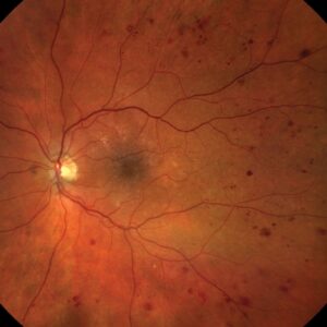
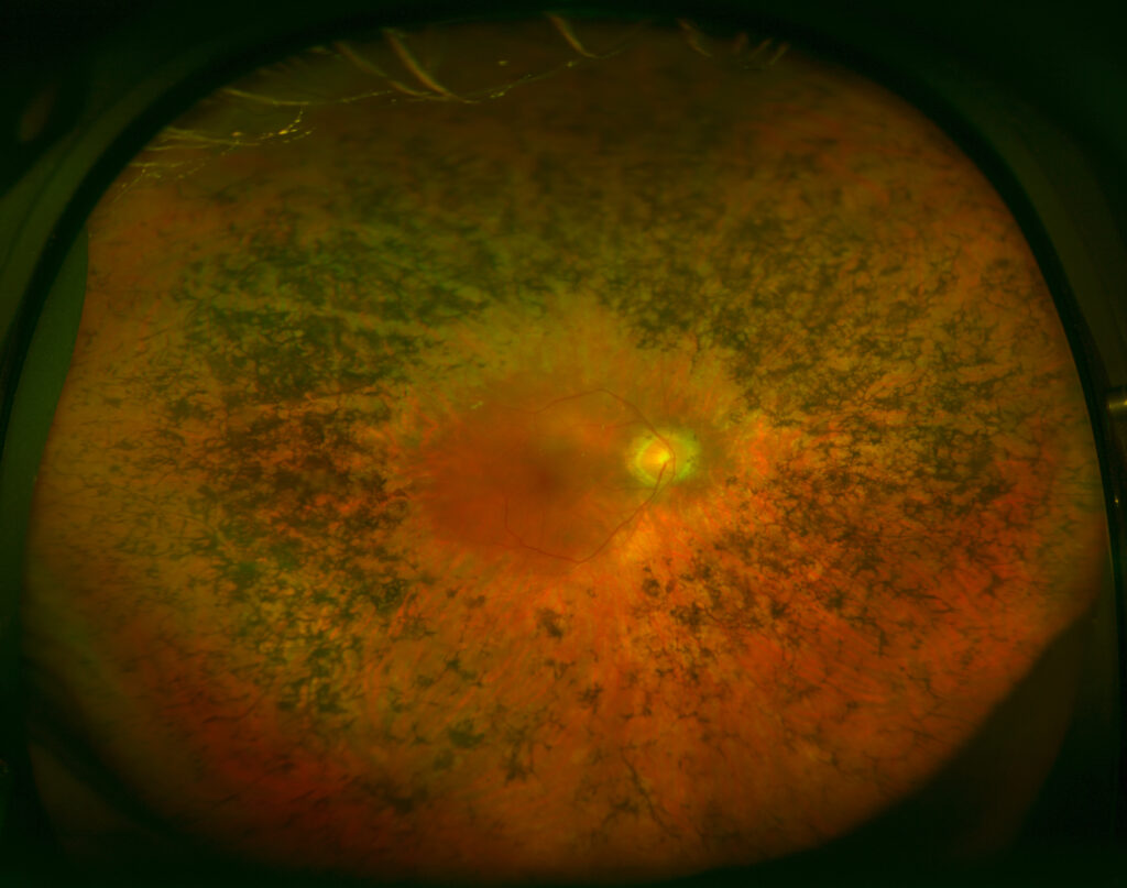
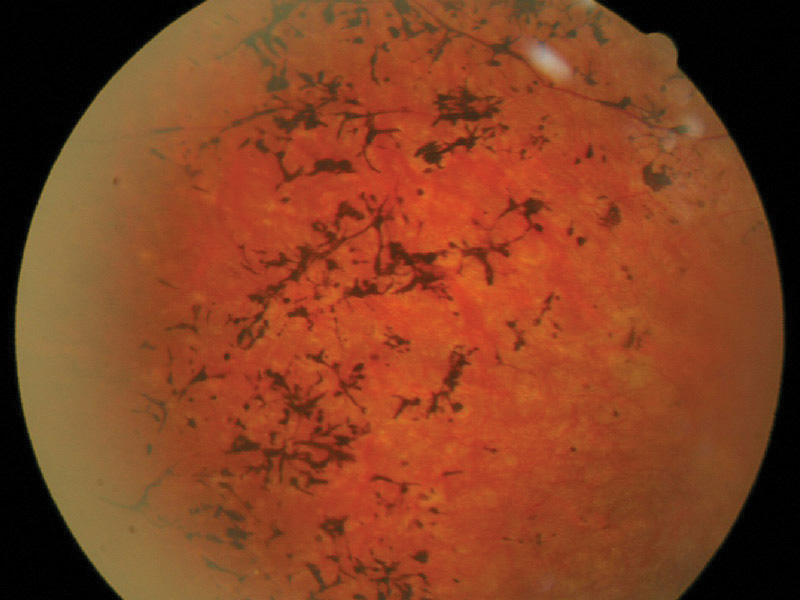
The earliest fundus change is often subtle arteriolar attenuation, and then fine pigment dusting or a granular, mottled RPE appears in the midperiphery. Early disc pallor is often absent, so a normal disc does not exclude disease. Finally, some patients never develop visible pigment, a picture called RP sine pigmento.
Vitreous and Lens Clues in RP Early Signs
Pigmented cells and fine dust in the anterior vitreous are common early findings, and posterior subcapsular cataract can also develop in young adults. Therefore, a young patient with PSC and night complaints deserves a dilated peripheral exam. These signs are easy to see at the slit lamp, even through a small pupil.
Fundus Autofluorescence in RP Early Signs
Autofluorescence often shows a hyperautofluorescent ring around the fovea, which marks the border between preserved and degenerating photoreceptors. In addition, mottled midperipheral hypoautofluorescence may precede visible atrophy, and over time the ring constricts, so serial imaging tracks progression.
OCT Findings in Early Disease
On OCT, the ellipsoid zone shortens from the periphery toward the fovea, and the outer nuclear layer thins in the same region while the inner retina stays relatively intact. Ellipsoid zone width therefore offers a reproducible marker (see ellipsoid zone disruption), and OCT also detects early cystoid spaces and epiretinal membrane.
Electroretinography and RP Early Signs
The full-field ERG is the main functional test, and rod responses decline first while cone responses fall later, with implicit times often delayed even when amplitudes look borderline. As a result, the ERG confirms disease while the fundus still looks near normal, and testing should follow ISCEV standards so results remain comparable over time.
Visual Field Testing
Goldmann kinetic perimetry shows early midperipheral defects well. However, central static programs may miss far peripheral loss, and you should also record baseline isopter areas because they guide later counseling.
Conditions That Mimic RP Early Signs
Several conditions cause night blindness or pigment that resembles early RP.
- Vitamin A deficiency after bariatric surgery or malabsorption
- Thioridazine toxicity and other drug retinopathies
- Syphilis, rubella and other infectious retinopathies
- Autoimmune retinopathy, including cancer-associated retinopathy
- Congenital stationary night blindness, which does not progress
- Unilateral pigment after trauma or old retinal detachment
When to Suspect Syndromic RP
Hearing loss suggests Usher syndrome, and similarly, obesity, polydactyly or renal disease point toward Bardet-Biedl syndrome. Therefore, refer patients with systemic features to genetics early.
Genetic Testing After RP Early Signs
Genetic testing now completes the workup in most retina centers, and a confirmed gene clarifies inheritance and recurrence risk for relatives. It also determines eligibility for gene therapy, for example voretigene for biallelic RPE65 disease, although a negative panel does not exclude RP.
Counseling at the First Visit
Explain that most forms progress slowly over decades, and discuss night driving, lighting at work and low vision services. Then, plan yearly review with OCT, autofluorescence and fields, and finally mention cataract and cystoid macular edema, because both are treatable.
Pitfalls to Avoid
- Do not dismiss night blindness because acuity is normal
- Never diagnose RP from the fundus alone without an ERG
- Do not miss treatable mimics such as vitamin A deficiency or syphilis
- Avoid relying on central static fields when peripheral loss is suspected


Document what you see
Two smartphone imaging tools built for everyday clinic use — one for the slit lamp, one for the fundus.
From Choroida — the team behind this siteReferences
- Hartong DT, Berson EL, Dryja TP. Retinitis pigmentosa. Lancet. 2006;368:1795-1809.
- Verbakel SK, van Huet RAC, Boon CJF, et al. Non-syndromic retinitis pigmentosa. Prog Retin Eye Res. 2018;66:157-186.
- Robson AG, Saihan Z, Jenkins SA, et al. Functional characterisation and serial imaging of abnormal fundus autofluorescence in patients with retinitis pigmentosa and normal visual acuity. Br J Ophthalmol. 2006;90:472-479.
- Robson AG, Frishman LJ, Grigg J, et al. ISCEV Standard for full-field clinical electroretinography (2022 update). Doc Ophthalmol. 2022;144:165-177.
- Hood DC, Lazow MA, Locke KG, Greenstein VC, Birch DG. The transition zone between healthy and diseased retina in patients with retinitis pigmentosa. Invest Ophthalmol Vis Sci. 2011;52:101-108.

Writes and reviews the clinical articles on Educate. Every post is checked against peer-reviewed sources before it goes live.