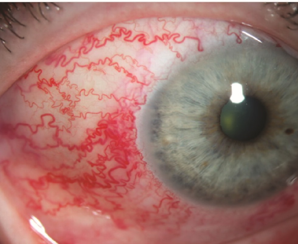
Corkscrew vessels are dilated, tortuous episcleral and conjunctival veins that carry arterial blood.
The pattern usually signals a carotid-cavernous fistula, which transmits arterial pressure into the orbital veins. However, patients often arrive labeled with chronic conjunctivitis or episcleritis, so a careful slit-lamp exam should separate these conditions early.
Why Corkscrew Vessels Form
In a fistula, arterial blood enters the cavernous sinus directly or through dural branches, then the superior ophthalmic vein reverses its flow and drains forward into the orbit, and as a result thin-walled episcleral veins dilate, lengthen and coil. The same pressure also raises episcleral venous pressure and, therefore, intraocular pressure.
Direct and Indirect Fistulas
Direct fistulas are high-flow shunts that usually follow head trauma, and they tend to cause abrupt chemosis, proptosis and an audible bruit. In contrast, indirect dural fistulas are low-flow and often arise spontaneously in older women, so their signs develop slowly and the red eye may persist for months before diagnosis. Barrow classified these shunts as types A to D according to arterial supply.
All-fit Slit-Lamp Adapter
Record and share exactly what you see at the slit lamp. One adapter fits any slit lamp or surgical microscope — and any smartphone.
From Choroida — the team behind this siteCorkscrew Vessels at the Slit Lamp
The vessels look engorged, dark red and spiral along their course, and they run radially and reach the limbus with nearly uniform caliber. They also lie deep and stay still when you move the conjunctiva with a cotton tip. Mild chemosis and lid fullness often accompany them, and for technique, see our slit-lamp exam guide.
What the Patient Reports
Many patients describe a red eye that never responds to drops, while others notice a whooshing noise in time with the pulse. Diplopia, a dull ache and blurred vision may also occur, but itching and sticky discharge are usually absent.
Corkscrew Vessels vs Conjunctivitis
Conjunctivitis produces diffuse injection that is most intense in the fornices and fades toward the limbus, and, in addition, discharge, follicles or papillae point toward infection or allergy. Corkscrew vessels, by contrast, form focal tortuous trunks that reach the cornea.
Corkscrew Vessels vs Episcleritis
Episcleritis causes sectoral or diffuse redness with mild tenderness, and its straight, radial vessels usually occupy one inflamed zone. Topical phenylephrine usually blanches this injection, which supports the diagnosis, while in contrast arterialized vessels typically stay engorged because pressure drives their dilation. Our episcleritis overview covers its typical course.
Tonometry in Corkscrew Vessels
First, measure intraocular pressure in both eyes, because a higher reading on the red side suggests raised episcleral venous pressure. The mires may also pulsate more because ocular pulse amplitude increases, and therefore pressure asymmetry in a so-called chronic conjunctivitis deserves further workup.
Gonioscopy Findings
Gonioscopy often shows blood in Schlemm canal, but excess lens pressure can also cause blood reflux, so use light contact. The angle usually remains open early in the course.
Bedside Signs That Raise Suspicion
- Unilateral red eye lasting weeks despite treatment
- Tortuous vessels that reach the limbus
- Higher intraocular pressure on the red side
- Blood in Schlemm canal on gonioscopy
- Proptosis, chemosis or a bruit over the closed lid
- Sixth nerve palsy or other ocular motor deficits
Fundus Findings
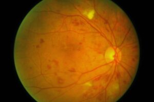
Dilated fundus examination may show dilated retinal veins and scattered hemorrhages, and choroidal folds or effusion can also appear in congested orbits. In severe cases, central retinal vein occlusion can threaten vision.
Imaging Corkscrew Vessels
Orbital CT or MRI may show an enlarged superior ophthalmic vein and swollen extraocular muscles, and color Doppler can also show reversed flow in the superior ophthalmic vein. Next, CT or MR angiography can suggest the shunt, but catheter angiography remains the reference standard for confirming and classifying the fistula.
Other Causes of Dilated Episcleral Vessels
Similar vessels can appear in several other conditions.
- Thyroid eye disease, with injection over the rectus insertions
- Sturge-Weber syndrome and episcleral hemangioma
- Orbital venous varix or superior vena cava obstruction
- Idiopathic elevated episcleral venous pressure
Management of Corkscrew Vessels
Treatment targets the shunt itself, and interventional neuroradiologists usually close direct fistulas with endovascular coils or other embolic agents. Many indirect fistulas need transvenous embolization, while some low-flow shunts close spontaneously, and meanwhile the ophthalmologist controls intraocular pressure, typically with aqueous suppressants. Tell patients that the vessels usually regress after closure, although this may take weeks, and our carotid-cavernous fistula article details these options.
Follow-up in Corkscrew Vessels
Recheck acuity, pressure, optic disc and fundus at each visit, since transient worsening after closure can occur, sometimes from venous thrombosis. However, cortical venous drainage on angiography raises stroke risk and needs prompt treatment, and redness may also return if the fistula reopens.
Pitfalls to Avoid
- Calling a unilateral chronic red eye conjunctivitis without measuring pressure
- Repeating antibiotic or steroid courses for persistent redness
- Assuming that a silent orbit excludes a fistula
- Pressing hard during gonioscopy and misreading blood reflux


Document what you see
Two smartphone imaging tools built for everyday clinic use — one for the slit lamp, one for the fundus.
From Choroida — the team behind this siteReferences
- Barrow DL, Spector RH, Braun IF, et al. Classification and treatment of spontaneous carotid-cavernous sinus fistulas. J Neurosurg. 1985;62:248-256.
- Miller NR. Diagnosis and management of dural carotid-cavernous sinus fistulas. Neurosurg Focus. 2007;23(5):E13.
- Ellis JA, Goldstein H, Connolly ES, Meyers PM. Carotid-cavernous fistulas. Neurosurg Focus. 2012;32(5):E9.
- Phelps CD, Thompson HS, Ossoinig KC. The diagnosis and prognosis of atypical carotid-cavernous fistula (red-eyed shunt syndrome). Am J Ophthalmol. 1982;93:423-436.
- Watson PG, Hayreh SS. Scleritis and episcleritis. Br J Ophthalmol. 1976;60:163-191.

Writes and reviews the clinical articles on Educate. Every post is checked against peer-reviewed sources before it goes live.