Macular hole vs ERM is a daily question in retina clinics, because both conditions cause central distortion in older adults.
Each one arises at the vitreomacular interface, but they differ in symptoms, bedside tests, OCT findings and surgical expectations.
Two Different Interface Problems
A full-thickness macular hole is a defect through every neural retinal layer at the fovea, while an epiretinal membrane is a fibrocellular sheet that grows on the inner retinal surface. The hole removes foveal tissue, while the membrane contracts and wrinkles it. As a result, the clinical picture and the prognosis follow different paths.
Who Gets Macular Hole vs ERM
Idiopathic macular holes typically affect women in their sixties and seventies, and they usually follow anomalous posterior vitreous detachment with focal foveal traction. ERM also rises with age and posterior vitreous detachment, and in addition it follows retinal surgery, retinal tears, vein occlusion, diabetic retinopathy and uveitis.
Fundus Explorer Pro
Photograph the retinal findings described here with the phone already in your pocket — 22 D optics and built-in illumination in one handheld unit.
From Choroida — the team behind this siteSymptoms in Macular Hole vs ERM
Patients with a hole often notice a central dark or missing spot, and straight lines may also bend inward toward the defect. ERM patients more often describe wavy lines, blur and size differences between the eyes.
Fundus Appearance
A hole looks like a round red defect at the fovea, so next, look for a gray cuff of subretinal fluid and small yellow deposits at its base. An ERM gives a glistening cellophane sheen with radiating striae, and advanced membranes also straighten and drag the perifoveal vessels.
The Watzke-Allen Test in Macular Hole vs ERM
First, project a thin vertical slit beam across the fovea with a contact or noncontact lens, and a patient with a full-thickness hole typically reports a break or gap in the line. With an ERM or a pseudohole, the line usually stays continuous, although it may look bent or thinned. However, this subjective sign has limited sensitivity, so OCT settles the question.
OCT Features of a Macular Hole
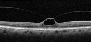
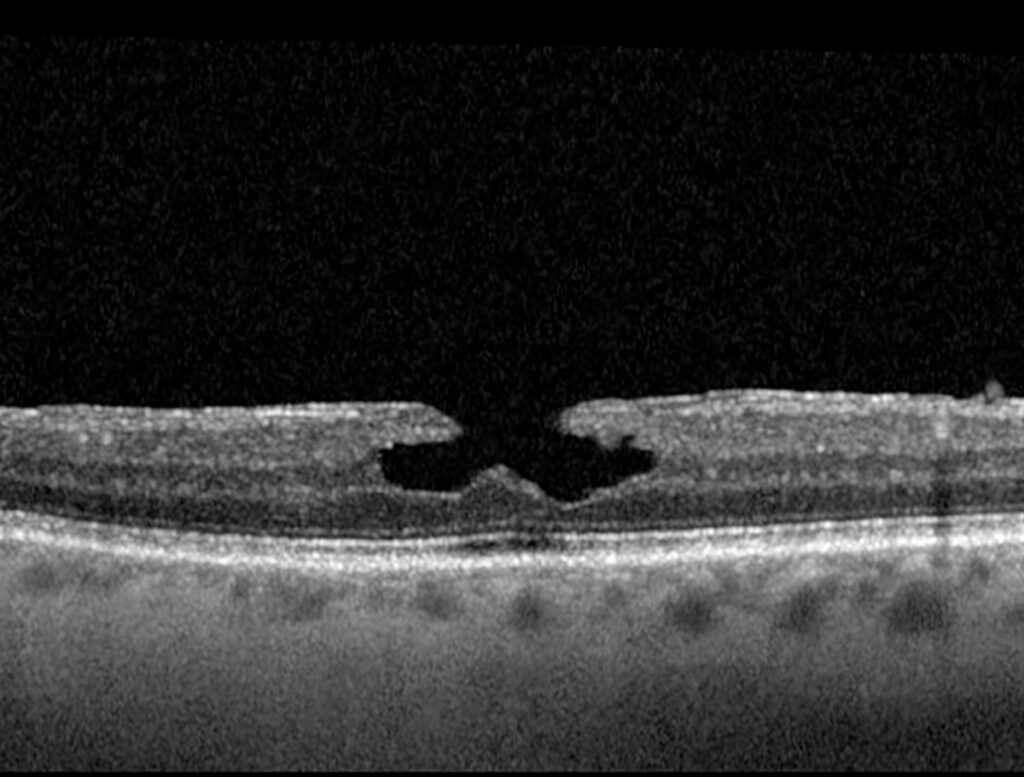
OCT shows a full-thickness foveal defect from the internal limiting membrane to the retinal pigment epithelium, and the edges are usually thickened with intraretinal cysts. The International Vitreomacular Traction Study classifies holes by minimum width, which then guides surgical planning (see macular hole staging and surgical timing).
OCT Features of an ERM
An ERM appears as a hyperreflective band on the inner retinal surface, and beneath it the foveal pit flattens and the retina thickens. Ectopic inner foveal layers suggest a more advanced stage and poorer visual prognosis. For separating ERM from traction, compare the patterns in ERM vs VMT on OCT.
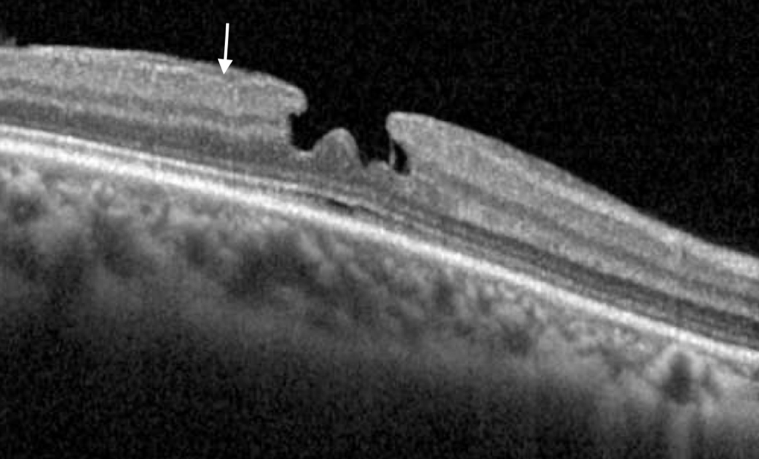
The Pseudohole Trap in Macular Hole vs ERM
A contracting membrane can steepen the foveal walls and mimic a round hole, and on fundus examination this pseudohole looks convincingly red. However, OCT shows preserved outer retina and photoreceptors at the base. Because the tissue remains, these eyes need membrane management, not hole surgery.
Natural History of Macular Hole vs ERM
Most established full-thickness holes persist or enlarge without treatment, although spontaneous closure occurs, mainly in small holes or after traction release. ERM, in contrast, often stays stable for long periods, so observation is reasonable while symptoms remain mild.
Surgery for a Macular Hole
Standard repair combines vitrectomy, internal limiting membrane peeling and gas tamponade, and anatomic closure rates are high in idiopathic holes, while large and chronic holes close less reliably. Also, warn phakic patients about cataract progression and flying restrictions with intraocular gas.
Surgery for an ERM
Vitrectomy with membrane peeling relieves traction on the inner retina, and many surgeons also peel the internal limiting membrane to reduce recurrence. Indications rest on symptoms, especially metamorphopsia, and on the patient’s visual demands.
Outcomes in Macular Hole vs ERM
After hole closure, vision improves gradually over many months, while ERM surgery often improves acuity, yet some distortion frequently persists. In addition, membranes can recur after peeling (see recurrence after peeling), and outer retinal integrity on OCT predicts final vision in both conditions.
Counseling the Patient
Explain that a hole is a gap, while a pucker is a scar-like layer. Next, set realistic goals: surgery aims to stabilize and improve vision, but rarely restores perfect sight. Finally, discuss gas, positioning, cataract and retinal detachment risks before consent.
Pitfalls to Avoid
- Do not diagnose a hole from a red fovea without OCT
- Never ignore the fellow eye, because it may show early traction
- Avoid relying on Watzke-Allen alone in poorly cooperative patients
- Do not promise complete resolution of distortion after peeling


Document what you see
Two smartphone imaging tools built for everyday clinic use — one for the slit lamp, one for the fundus.
From Choroida — the team behind this siteReferences
- Duker JS, Kaiser PK, Binder S, et al. The International Vitreomacular Traction Study Group classification of vitreomacular adhesion, traction, and macular hole. Ophthalmology. 2013;120:2611-2619.
- Gass JD. Reappraisal of biomicroscopic classification of stages of development of a macular hole. Am J Ophthalmol. 1995;119:752-759.
- Watzke RC, Allen L. Subjective slitbeam sign for macular disease. Am J Ophthalmol. 1969;68:449-453.
- Govetto A, Lalane RA, Sarraf D, Figueroa MS, Hubschman JP. Insights into epiretinal membranes: presence of ectopic inner foveal layers and a new optical coherence tomography staging scheme. Am J Ophthalmol. 2017;175:99-113.
- Kelly NE, Wendel RT. Vitreous surgery for idiopathic macular holes: results of a pilot study. Arch Ophthalmol. 1991;109:654-659.
Test yourself
A few questions straight from this article.
-
How is a full-thickness macular hole defined, in contrast to an epiretinal membrane?
A full-thickness hole removes foveal tissue through every neural layer, whereas an ERM is a fibrocellular sheet that contracts and wrinkles the inner retina. -
Which patients typically develop idiopathic full-thickness macular holes?
Idiopathic macular holes typically affect women in their sixties and seventies, usually after anomalous posterior vitreous detachment with focal foveal traction. -
Which symptom points more toward a macular hole than an epiretinal membrane?
Hole patients often notice a central dark or missing spot, while ERM patients more often describe wavy lines, blur and size differences between the eyes. -
What is the typical fundus appearance of an epiretinal membrane?
An ERM gives a glistening cellophane sheen with radiating striae, and advanced membranes straighten and drag the perifoveal vessels. -
On the Watzke-Allen test, what does a patient with a full-thickness macular hole typically report?
With a full-thickness hole the vertical slit beam typically appears broken; with an ERM or pseudohole it usually stays continuous, though the test has limited sensitivity and OCT settles the question. -
Which OCT feature of an epiretinal membrane suggests a more advanced stage and poorer visual prognosis?
ERM shows as an inner hyperreflective band with foveal flattening and thickening; ectopic inner foveal layers indicate a more advanced stage and poorer visual prognosis. -
What OCT finding identifies a macular pseudohole caused by a contracting epiretinal membrane?
A pseudohole can look convincingly red on fundus examination, but OCT shows preserved outer retina at its base, so these eyes need membrane management rather than hole surgery. -
By which measurement does the International Vitreomacular Traction Study classify full-thickness macular holes?
The International Vitreomacular Traction Study classifies holes by minimum width on OCT, and this guides surgical planning. -
What is the standard surgical repair for a full-thickness macular hole?
Standard repair combines vitrectomy, internal limiting membrane peeling and gas tamponade; closure rates are high in idiopathic holes, while large and chronic holes close less reliably. -
Which OCT finding predicts final vision after surgery for both macular hole and epiretinal membrane?
Outer retinal integrity on OCT predicts final vision in both conditions; vision improves gradually after hole closure, while some distortion often persists after ERM peeling.

Writes and reviews the clinical articles on Educate. Every post is checked against peer-reviewed sources before it goes live.