Mucous plaque keratopathy is a corneal complication of herpes zoster ophthalmicus.
It usually appears weeks to months after the acute rash, and the plaques look gray, elevated and oddly shaped on the epithelium. Because they can mimic dendrites, they often trigger an unnecessary change in antiviral therapy.
What Is Mucous Plaque Keratopathy?
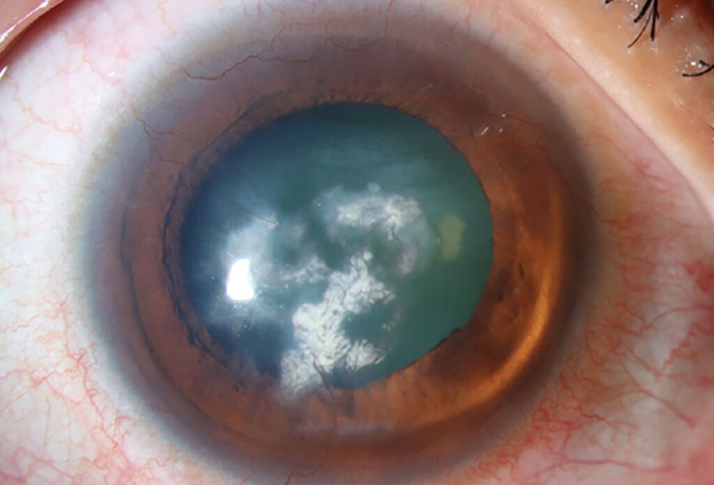
Mucous plaque keratopathy consists of mucus adherent to an abnormal corneal epithelium, and the plaques sit on the surface and lack the swollen epithelial cells of an active dendrite. Marsh’s early descriptions of zoster keratitis also included these late surface lesions.
When It Appears After Zoster
Timing helps the diagnosis, because pseudodendrites usually appear early, within the first days to weeks. In contrast, mucous plaques tend to appear later, often months after the rash. Therefore, always ask when the shingles started (see herpes zoster ophthalmicus).
All-fit Slit-Lamp Adapter
Record and share exactly what you see at the slit lamp. One adapter fits any slit lamp or surgical microscope — and any smartphone.
From Choroida — the team behind this siteSlit Lamp Signs of Mucous Plaque Keratopathy
- Gray-white, elevated plaques with sharp borders
- Linear, branching or irregular shapes
- Loose adherence, so a cotton tip lifts them easily
- Shape and position that change between visits
- Brilliant staining with rose bengal or lissamine green
- Weaker staining with fluorescein
Why the Plaques Form
The mechanism is not fully settled, but most authors link the plaques to reduced corneal sensation and an unstable tear film. In addition, chronic inflammation may alter the epithelial surface and its mucus layer, and some reports have detected viral material in plaques, although its role remains uncertain.
Associated Findings
Mucous plaques rarely occur alone, so look for stromal keratitis, endotheliitis and anterior uveitis in the same eye. Also check intraocular pressure, because zoster trabeculitis can raise it, and finally test corneal sensation before you instill anesthetic. Symptoms are often mild, while the surface disease may look severe.
Mucous Plaque Keratopathy vs Pseudodendrites
Pseudodendrites are elevated epithelial lesions made of swollen cells that lack the terminal bulbs of herpes simplex dendrites (see pseudodendrite morphology). Mucous plaques also lack terminal bulbs, but they wipe off easily and shift over time. In addition, pseudodendrites belong to the early phase, while plaques belong to the late phase.
Mucous Plaque Keratopathy vs Filaments
Filaments are strands of mucus and epithelium attached at one end to the cornea, and they move with the blink and often cause marked pain. Mucous plaques, however, lie flat and broad on the surface, but both reflect a damaged surface, so they can coexist in the same eye.
Ruling Out HSV
A branching lesion in a zoster patient still raises the question of herpes simplex, but true HSV dendrites show terminal bulbs and central ulceration that stains with fluorescein. Therefore, look carefully at the lesion ends and the staining pattern, and when doubt remains, a PCR swab can help.
Other Conditions to Consider
Exposure keratopathy and toxic epitheliopathy can also leave mucus on the surface. Therefore, review the drop history and lid closure at every visit. Epithelial basement membrane changes may also mimic fine linear plaques, so examine the fellow eye.
Diagnosing Mucous Plaque Keratopathy
Diagnosis is clinical, so first confirm the zoster history and the timing. Next, stain with fluorescein and then with lissamine green, then gently touch the plaque with a cotton tip to test adherence.
Examination Pearls
First, instill fluorescein and wait for the dye to spread across the surface, then view the cornea with the cobalt blue filter and a green filter. Also, lissamine green highlights plaques that fluorescein can miss, so use both dyes before you commit to a diagnosis.
Treating Mucous Plaque Keratopathy
Management targets the surface and the inflammation, and preservative-free lubricants form the base of therapy. Topical acetylcysteine can dissolve the mucus, although evidence comes mainly from case series, and gentle debridement may also help, but plaques often recur.
Role of Steroids and Antivirals
Many clinicians use low-dose topical steroids when stromal or anterior chamber inflammation coexists, so taper slowly, because zoster inflammation tends to rebound. Some clinicians also add antivirals, given reports of viral material in plaques. However, no controlled trial has defined the best regimen.
Managing the Neurotrophic Component
Reduced sensation drives much of the chronic surface damage. For example, persistent epithelial defects may follow if the surface is neglected. In these cases, treat the cornea as neurotrophic (see neurotrophic keratitis staging), and options then include punctal plugs, bandage lenses and tarsorrhaphy.
Counseling the Patient
Explain that the plaques are a surface reaction after shingles and that they can return over months. Patients should report pain, redness or a drop in vision promptly. In addition, mention that vaccination helps prevent zoster in eligible adults.
Follow-Up in Mucous Plaque Keratopathy
Review the eye every few weeks during active disease, and at each visit record plaque location, sensation and pressure. Then, adjust steroids according to stromal and anterior chamber activity. Plan long-term follow-up, because zoster eye disease often runs a chronic course.
Pitfalls to Avoid
- Do not label every branching zoster lesion as HSV
- Never skip corneal sensation testing
- Avoid stopping steroids abruptly
- Avoid preserved drops in a neurotrophic cornea


Document what you see
Two smartphone imaging tools built for everyday clinic use — one for the slit lamp, one for the fundus.
From Choroida — the team behind this siteReferences
- Liesegang TJ. Herpes zoster ophthalmicus natural history, risk factors, clinical presentation, and morbidity. Ophthalmology. 2008;115(2 Suppl):S3-S12.
- Piebenga LW, Laibson PR. Dendritic lesions in herpes zoster ophthalmicus. Arch Ophthalmol. 1973;90:268-270.
- Marsh RJ. Herpes zoster keratitis. Trans Ophthalmol Soc U K. 1973;93:181-192.
- American Academy of Ophthalmology. Basic and Clinical Science Course, Section 8: External Disease and Cornea. San Francisco: AAO; 2023.
- Bowling B. Kanski’s Clinical Ophthalmology: A Systematic Approach. 8th ed. Elsevier; 2016.
Test yourself
A few questions straight from this article.
-
What is the composition of mucous plaque keratopathy in herpes zoster ophthalmicus?
Mucous plaque keratopathy consists of mucus adherent to an abnormal corneal epithelium; the plaques sit on the surface and lack the swollen epithelial cells of an active dendrite. -
When do mucous plaques typically appear in herpes zoster ophthalmicus, compared with pseudodendrites?
Pseudodendrites usually appear within the first days to weeks, while mucous plaques tend to appear later, often months after the rash, so the shingles onset date aids diagnosis. -
Which staining pattern is typical of zoster mucous plaques?
Mucous plaques stain brilliantly with rose bengal or lissamine green but more weakly with fluorescein, so both dyes should be used before committing to the diagnosis. -
How do zoster mucous plaques respond to gentle touch with a cotton tip?
Mucous plaques are loosely adherent, so a cotton tip lifts them easily, and their shape and position change between visits. -
Which factors do most authors link to the formation of mucous plaques after zoster?
The mechanism is not fully settled, but most authors link mucous plaques to reduced corneal sensation and an unstable tear film, with chronic inflammation altering the surface. -
Which features identify a true herpes simplex dendrite in a zoster patient with a branching corneal lesion?
True HSV dendrites show terminal bulbs and central ulceration that stains with fluorescein, so the lesion ends and staining pattern are checked, with a PCR swab if doubt remains. -
How do corneal filaments differ from mucous plaques in zoster keratopathy?
Filaments are strands of mucus and epithelium attached at one end that move with the blink and often hurt, whereas plaques lie flat and broad; both can coexist. -
Which topical agent can dissolve the mucus in mucous plaque keratopathy?
Preservative-free lubricants form the base of therapy, and topical acetylcysteine can dissolve the mucus, although evidence comes mainly from case series and plaques often recur. -
Why are topical steroids tapered slowly when used for inflammation accompanying mucous plaque keratopathy?
Low-dose topical steroids are used when stromal or anterior chamber inflammation coexists, and they are tapered slowly because zoster inflammation tends to rebound. -
Why should intraocular pressure be checked in an eye with zoster mucous plaques?
Mucous plaques rarely occur alone, so the eye is checked for stromal keratitis, endotheliitis and uveitis, and the pressure is measured because zoster trabeculitis can raise it.

Writes and reviews the clinical articles on Educate. Every post is checked against peer-reviewed sources before it goes live.