Eyelid melanoma is a malignant tumor of melanocytes arising in the eyelid skin or at the lid margin.
It is rare among eyelid cancers, yet it causes a disproportionate share of tumor deaths. Therefore, every new or changing pigmented lid lesion deserves a careful slit lamp assessment.
What Eyelid Melanoma Is
Primary disease starts in epidermal melanocytes of the lid skin or the mucocutaneous junction, and a conjunctival melanoma can also spread onto the margin and mimic a skin primary. Because the margin joins skin and mucosa, one tumor can behave like either disease.
Who Is at Risk
Most patients are older adults with fair skin and long sun exposure, so also ask about prior melanoma, family history, multiple atypical nevi and immunosuppression. However, melanoma occurs in every skin type, so a low-risk profile never excludes it.
All-fit Slit-Lamp Adapter
Record and share exactly what you see at the slit lamp. One adapter fits any slit lamp or surgical microscope — and any smartphone.
From Choroida — the team behind this siteClinical Subtypes of Eyelid Melanoma
- Lentigo maligna melanoma: a flat, slowly enlarging brown patch on sun-damaged skin
- Superficial spreading melanoma: a flat or slightly raised lesion with mixed colors
- Nodular melanoma: a dome-shaped dark nodule that grows quickly
- Amelanotic melanoma: a pink lesion that easily passes for a benign bump
History and Slit Lamp Examination
Ask when the spot appeared and whether it has grown, darkened, itched or bled. Then examine the lesion at low and high magnification, assessing border, surface and color. Finally, palpate the preauricular and submandibular nodes, which drain the eyelids.
Lid Margin Signs of Eyelid Melanoma
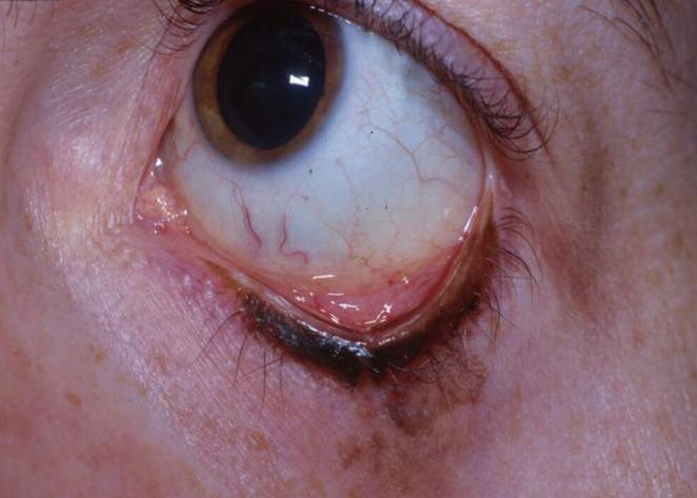
In general, malignant lesions destroy normal margin architecture, so look for lash loss, notching, distorted meibomian orifices and ulceration. In contrast, a benign margin nevus usually keeps its lashes and smooth surface (see eyelid nevus). Irregular pigment streaking along the margin is another warning sign.
Everting the Lid
A small margin lesion may hide a larger posterior component, so evert the lid in every case and inspect the tarsal conjunctiva and fornix. Flat pigment, nodules or feeder vessels on the palpebral surface suggest spread.
Conjunctival Extension in Eyelid Melanoma
Conjunctival spread changes both prognosis and treatment, and published series suggest that margin and conjunctival involvement carry a higher risk of local recurrence. Next, check the bulbar conjunctiva, caruncle and plica for primary acquired melanosis, then manage that component with the principles used for conjunctival melanoma.
Benign Mimics of Eyelid Melanoma
- Intradermal or compound nevus
- Seborrheic keratosis
- Pigmented basal cell carcinoma
- Solar lentigo
- Apocrine hidrocystoma with a bluish hue
Biopsy Triggers for Eyelid Melanoma
- A new pigmented lesion in an older adult
- Documented growth or color change
- Lash loss or margin notching over the lesion
- Ulceration, bleeding or crusting
- Irregular border or several shades of color
- Pigment extending onto the tarsal or bulbar conjunctiva
Biopsy Technique
Small lesions with simple closure suit excisional biopsy with narrow margins, while for larger lesions you should take a punch or incisional biopsy from the thickest area. Avoid shave biopsy, because transection prevents accurate Breslow thickness measurement.
Staging Eyelid Melanoma
Eyelid skin melanoma follows the AJCC staging system for cutaneous melanoma, and Breslow thickness and ulceration drive the T category and prognosis. As a result, the pathology report determines excision margins and nodal assessment.
Surgical Management
Complete excision with histologically clear margins is the primary treatment, but wide cutaneous margins are hard near the eye, so surgeons balance clearance with lid function. Staged excision with permanent sections, or Mohs surgery with immunostains, confirms clearance while sparing tissue. Delay reconstruction until margins are clear (see eyelid margin reconstruction).
Treating the Conjunctival Component
Surgeons excise conjunctival disease with a no-touch technique and often add cryotherapy to the edges. Topical mitomycin C may also treat residual primary acquired melanosis, but drops cannot replace complete excision of an invasive tumor.
Nodes and Systemic Therapy
Sentinel lymph node biopsy is often considered for thicker or ulcerated tumors, and it refines staging, while its survival benefit remains debated. Patients with nodal or distant disease need a melanoma oncology team for checkpoint inhibitors or targeted therapy.
Follow-Up After Eyelid Melanoma
Recurrence can appear years after surgery, so examine the scar, margin, conjunctiva and regional nodes at each visit, and photograph the area. Patients also need full skin checks by a dermatologist, because second primaries occur.
Counseling in Eyelid Melanoma
Explain that only a biopsy confirms the diagnosis, and describe the surgery, reconstruction and expected lid appearance in plain terms. Also teach sun protection and regular mirror checks of the lid, and finally list the new signs that should prompt an early visit.
Pitfalls to Avoid
- Do not label a margin lesion a nevus before everting the lid
- Never shave a suspicious pigmented lesion
- Do not dismiss a pink nodule, because amelanotic melanoma exists
- Avoid closing the defect before margins are confirmed


Document what you see
Two smartphone imaging tools built for everyday clinic use — one for the slit lamp, one for the fundus.
From Choroida — the team behind this siteReferences
- Cook BE Jr, Bartley GB. Epidemiologic characteristics and clinical course of patients with malignant eyelid tumors in an incidence cohort in Olmsted County, Minnesota. Ophthalmology. 1999;106:746-750.
- Esmaeli B, Wang B, Deavers M, et al. Prognostic factors for survival in malignant melanoma of the eyelid skin. Ophthalmic Plast Reconstr Surg. 2000;16:250-257.
- Shields CL, Shields JA, Gündüz K, et al. Conjunctival melanoma: risk factors for recurrence, exenteration, metastasis, and death in 150 consecutive patients. Arch Ophthalmol. 2000;118:1497-1507.
- Gershenwald JE, Scolyer RA, Hess KR, et al. Melanoma staging: evidence-based changes in the American Joint Committee on Cancer eighth edition cancer staging manual. CA Cancer J Clin. 2017;67:472-492.

Writes and reviews the clinical articles on Educate. Every post is checked against peer-reviewed sources before it goes live.