Palisades of Vogt are radial fibrovascular ridges at the corneoscleral limbus.
They mark the niche where limbal epithelial stem cells live. Therefore, a careful look at them tells you a lot about how well the cornea can repair itself.
What Are the Palisades of Vogt?
The palisades of Vogt are stromal folds that run perpendicular to the corneal edge, and between the ridges the epithelium dips into deep rete pegs and crypts. Each ridge carries a fibrovascular core with a fine radial vessel, so the healthy limbus looks finely striated, like a short picket fence.
Stem Cells Within the Palisades of Vogt
Basal cells between the ridges divide slowly and behave as stem cells, and Cotsarelis and colleagues identified these slow-cycling, label-retaining basal cells at the limbus. Later, Dua and colleagues described limbal epithelial crypts extending from the palisades, and the ridged shape protects these cells and keeps blood supply close to them.
All-fit Slit-Lamp Adapter
Record and share exactly what you see at the slit lamp. One adapter fits any slit lamp or surgical microscope — and any smartphone.
From Choroida — the team behind this siteWhere to Look at the Limbus
The palisades are usually clearest at the superior and inferior limbus, because these zones sit under the lids, which shield them from light and trauma. In contrast, the nasal and temporal limbus often shows fewer, shorter ridges. Therefore, lift the upper lid and pull down the lower lid before you call them absent.
Slit Lamp Technique for Palisades of Vogt
First, survey the whole limbus with diffuse illumination and low magnification, and next raise the magnification and narrow the beam. Then place the beam just inside the limbus, so scattered light backlights the ridges. Sclerotic scatter and retroillumination from the iris also outline them well, and for beam settings in detail, see slit lamp illumination techniques.
Pigmentation and Palisades of Vogt
Melanocytes sit in the limbal basal epithelium between the ridges, so patients with darker skin often show brown, clearly visible palisades. In lightly pigmented eyes, the ridges look translucent and gray, which makes them easy to miss without careful focusing.
How Age Changes the Picture
Palisades are usually most distinct in young eyes, but with age, they often become flatter, fewer and less regular. Therefore, faint palisades in an older patient with a healthy cornea need no treatment.
Fluorescein and Imaging
Fluorescein collects in the grooves between ridges and accentuates the pattern, while in vivo confocal microscopy shows palisades, crypts and basal cells at cellular resolution. Anterior segment OCT can also display the undulating limbal epithelium in cross section.
Loss of Palisades of Vogt in LSCD
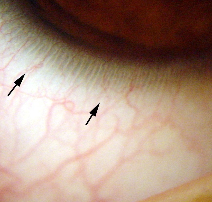
Limbal stem cell deficiency depletes or destroys the stem cell niche, so the palisades flatten or vanish over the affected clock hours. The global consensus lists loss of palisades among the clinical signs of LSCD. However, the diagnosis rests on the whole picture, and impression cytology or confocal microscopy can confirm it (see LSCD classification).
Signs That Travel With Palisade Loss
Look for these companion signs:
- Dull, irregular, thin epithelium over the affected sector
- Late fluorescein staining in a whorl or vortex pattern
- Superficial vessels crossing the limbus onto the cornea
- Conjunctival epithelium growing onto the cornea, then pannus
- Recurrent or persistent epithelial defects
Common Causes of Limbal Damage
Chemical and thermal burns cause the most dramatic and extensive loss, while aniridia, Stevens-Johnson syndrome and mucous membrane pemphigoid can also deplete the limbus. Iatrogenic causes include multiple limbal surgeries, cryotherapy and topical antimetabolites such as mitomycin C, and long-term contact lens wear can produce a slower, sectoral pattern.
Palisades of Vogt in Surgical Planning
Before conjunctival limbal autograft, surgeons examine the fellow eye’s limbus closely, and a healthy superior limbus with clear palisades suggests a reasonable donor site. However, visible ridges do not prove an adequate stem cell reserve, and in partial deficiency, intact sectors of the same eye also matter, because they still supply epithelium. Harvesting too much tissue can induce deficiency in the donor eye.
Contact Lens Wear and the Limbus
Chronic soft lens wear, especially with poor fit or low oxygen transmission, can stress the superior limbus, and early clues include superior epithelial irregularity and blurred palisade detail. Therefore, inspect the superior limbus at every lens review, and when the patient stops wear and you treat the surface, mild changes may recover.
Counseling the Patient
Most patients never hear of the palisades until a problem appears, so explain that the limbus holds the cells that renew the corneal surface. Then link the finding to the plan, whether monitoring, a lens holiday or referral. For example, serial photographs help patients see stability or change.
Pitfalls With the Palisades of Vogt
- Calling palisades absent without lifting the lids
- Treating faint palisades in an elderly eye as disease
- Confusing them with the white limbal girdle of Vogt
- Diagnosing LSCD from palisade loss alone
- Mistaking normal pigmented palisades for an acquired pigmented lesion


Document what you see
Two smartphone imaging tools built for everyday clinic use — one for the slit lamp, one for the fundus.
From Choroida — the team behind this siteReferences
- Goldberg MF, Bron AJ. Limbal palisades of Vogt. Trans Am Ophthalmol Soc. 1982;80:155-171.
- Cotsarelis G, Cheng SZ, Dong G, Sun TT, Lavker RM. Existence of slow-cycling limbal epithelial basal cells that can be preferentially stimulated to proliferate: implications on epithelial stem cells. Cell. 1989;57:201-209.
- Dua HS, Shanmuganathan VA, Powell-Richards AO, Tighe PJ, Joseph A. Limbal epithelial crypts: a novel anatomical structure and a putative limbal stem cell niche. Br J Ophthalmol. 2005;89:529-532.
- Deng SX, Borderie V, Chan CC, et al. Global consensus on definition, classification, diagnosis, and staging of limbal stem cell deficiency. Cornea. 2019;38:364-375.

Writes and reviews the clinical articles on Educate. Every post is checked against peer-reviewed sources before it goes live.