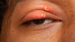
Foreign body sensation is one of the commonest complaints in eye clinics, and often no foreign body is found.
The symptom arises from irritation of corneal and conjunctival nerve endings. Therefore, the cause usually lies on the ocular surface, the lid margin or the tarsal conjunctiva. A fixed examination order finds most causes within a few minutes.
What the Patient Means by Foreign Body Sensation
Patients describe sand, grit, an eyelash or something stuck in the eye, but timing and location help more than the exact words. Sudden onset after wind, grinding or gardening suggests a true particle, whereas gradual bilateral grittiness points to a surface disorder.
History in Foreign Body Sensation
First, ask about onset, laterality and contact lens wear. Pain on every blink suggests a lesion under the upper lid, and morning symptoms suggest exposure or floppy lids, while evening symptoms suggest dry eye.
All-fit Slit-Lamp Adapter
Record and share exactly what you see at the slit lamp. One adapter fits any slit lamp or surgical microscope — and any smartphone.
From Choroida — the team behind this siteStart With the Lids and Lashes
Examine the lids under low magnification before touching the eye, and look at lid position, blink completeness and margin disease.
Then check every lash along both margins, including the lateral canthus. A single misdirected lash can cause intense symptoms (see trichiasis).
Distichiasis and Fine Lashes
Distichiatic lashes emerge from meibomian gland orifices and are often thin and pale, so diffuse light misses them. Therefore, use high magnification and retroillumination along the posterior margin.
Evert the Upper Lid in Foreign Body Sensation
Upper lid eversion is the step most often skipped. However, a subtarsal particle classically leaves vertical linear corneal abrasions.
Evert the lid and inspect the tarsal plate with fluorescein (see subtarsal foreign body), and sweep the upper fornix with a moistened swab when the history is strong.
Fluorescein Staining Patterns
Fluorescein turns a vague complaint into a map. For example, inferior punctate staining suggests exposure. Interpalpebral staining fits aqueous deficient dry eye, while superior staining suggests lid or limbal disease.
Dry Eye as a Cause of Foreign Body Sensation
Dry eye is the commonest cause when the cornea looks almost normal. Measure tear breakup time, then stain with lissamine green and inspect meibomian gland orifices and expressed secretions. Symptoms and signs often disagree, so a mild exam does not exclude dry eye.
Filaments on the Cornea
Filaments are strands of mucus and epithelium attached to the cornea, and they tug with each blink, so patients report sharp recurring grit.
Look for them with fluorescein, often on the upper cornea. Treat the underlying surface disease, because removal alone gives brief relief (see filamentary keratitis).
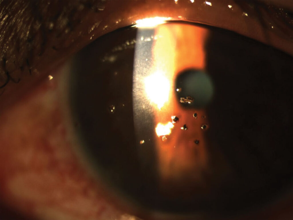
Superior Limbic Keratoconjunctivitis
SLK causes marked grit with surprisingly little visible redness. Ask the patient to look down while you lift the upper lid, then look for sectoral superior bulbar injection, thickening and punctate staining. Consider thyroid testing as well, because SLK frequently accompanies thyroid eye disease.
Conjunctivochalasis
Redundant bulbar conjunctiva folds over the lower lid margin. As a result, it disturbs the tear meniscus and rubs during downgaze. Symptoms often worsen when the patient looks down to read.
Floppy Eyelid and Exposure
A lax upper lid everts easily during sleep. Therefore, the patient wakes with irritation, mucus and a papillary reaction.
Pull the upper lid upward and note how easily it everts. Then ask about snoring, because floppy eyelid syndrome associates with obstructive sleep apnea.
Corneal Epithelial Causes of Foreign Body Sensation
Recurrent erosion typically causes sharp pain on waking. Look for map, dot or fingerprint lines and negative fluorescein staining.
Consider Thygeson superficial punctate keratitis when raised gray dots appear in a quiet eye, and check contact lens fit and deposits.
Drop Toxicity and Foreign Body Sensation
Chronic drops can cause the symptom by themselves. For example, preserved glaucoma drops often produce diffuse punctate staining.
Therefore, review every drop, including over the counter products. Switching to preservative free formulations often helps over several weeks.
Foreign Body Sensation With a Normal Exam
Some patients have intense symptoms with a quiet surface. In that case, consider neuropathic ocular pain, especially after refractive surgery.
Pain that persists after a topical anesthetic drop suggests a neuropathic component. Then repeat the exam at another visit, since signs fluctuate.
Counseling and Follow-up
Show patients their staining pattern, because a visible sign improves acceptance of a chronic diagnosis. Set a review date to confirm that treatment worked, and finally give clear return advice for pain, discharge or reduced vision.
Pitfalls to Avoid
- Do not skip upper lid eversion when linear abrasions appear
- Do not miss distichiasis by examining only with diffuse light
- Avoid labeling every case dry eye before checking lids and limbus
- Never give topical anesthetic drops for home use
- Do not forget SLK when superior staining dominates


Document what you see
Two smartphone imaging tools built for everyday clinic use — one for the slit lamp, one for the fundus.
From Choroida — the team behind this siteReferences
- Bowling B. Kanski’s Clinical Ophthalmology: A Systematic Approach. 8th ed. Edinburgh: Elsevier; 2016.
- Craig JP, Nichols KK, Akpek EK, et al. TFOS DEWS II definition and classification report. Ocul Surf. 2017;15:276-283.
- Nelson JD. Superior limbic keratoconjunctivitis (SLK). Eye (Lond). 1989;3:180-189.
- Meller D, Tseng SC. Conjunctivochalasis: literature review and possible pathophysiology. Surv Ophthalmol. 1998;43:225-232.
- Culbertson WW, Ostler HB. The floppy eyelid syndrome. Am J Ophthalmol. 1981;92:568-575.

Writes and reviews the clinical articles on Educate. Every post is checked against peer-reviewed sources before it goes live.