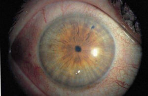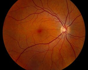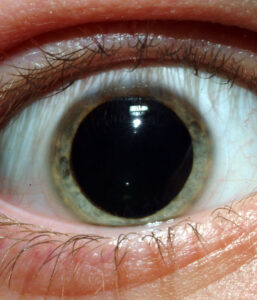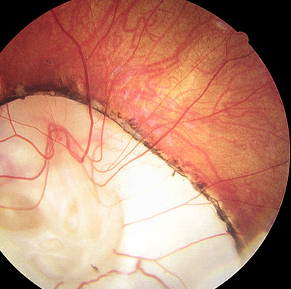
Optic disc anomalies are congenital or developmental variations in the size, shape or structure of the nerve head.
Most are found incidentally on routine fundus examination.
However, several carry retinal or systemic consequences.
Therefore, naming the anomaly correctly guides the workup.
Why Optic Disc Anomalies Matter
An unusual disc can mimic glaucoma, papilledema or a mass.
As a result, patients may undergo needless scans or treatment.
In addition, some anomalies predispose to macular or retinal detachment.
Others point to brain or endocrine malformations that need early care.
A Comparison of Optic Disc Anomalies
Use this summary as a quick bedside reference.
- Tilted disc: oval, oblique disc with inferonasal crescent; myopic astigmatism and superotemporal field loss
- Optic disc pit: gray oval depression, usually temporal; risk of maculopathy
- Coloboma: white excavation, deeper inferiorly; detachment and syndromic links
- Morning glory disc: enlarged funnel with central glial tuft; detachment and encephalocele risk
- Hypoplasia: small disc with double ring sign; pituitary and midline anomalies
- Megalopapilla: large disc and cup with healthy rim; usually normal function
- Drusen: lumpy, elevated disc without hyperemia; field loss and pseudopapilledema
Fundus Explorer Pro
Photograph the retinal findings described here with the phone already in your pocket — 22 D optics and built-in illumination in one handheld unit.
From Choroida — the team behind this siteTilted Disc
The nerve enters the globe obliquely, so the disc looks oval and rotated.
Vessels often emerge nasally, a pattern called situs inversus.
Also, an inferior staphyloma may cause superotemporal field defects that cross the vertical midline.
Correcting the refraction often reduces these defects, which helps separate them from chiasmal disease.
Optic Disc Pit
A pit appears as a small gray depression, most often at the temporal rim.
Vision stays normal until fluid tracks into the macula.
Then OCT shows schisis and serous detachment without a retinal break (see optic disc pit maculopathy).
Therefore, advise patients to report new blur or distortion promptly.
Optic Disc Coloboma
Incomplete closure of the optic fissure leaves a bowl-shaped white excavation.
The defect is usually deeper inferiorly and may extend into the choroid.
Because tissue is thin at the margin, retinal detachment can follow.
Also, consider CHARGE and renal coloboma syndromes in bilateral cases.
Morning Glory Disc
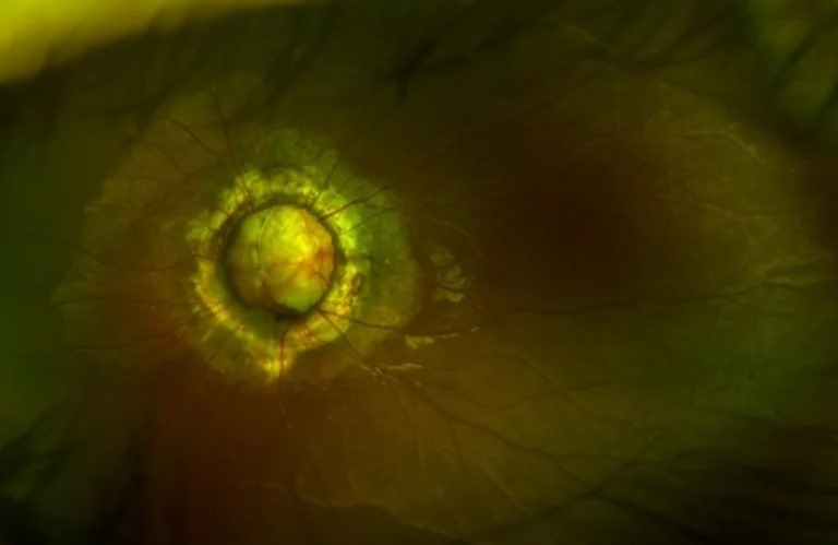
This anomaly shows an enlarged funnel-shaped disc with a central white glial tuft.
Straightened vessels radiate from the edge, surrounded by a pigment ring.
Most cases are unilateral, and vision is usually reduced.
In addition, retinal detachment may develop later in life.
Optic Nerve Hypoplasia
A hypoplastic disc looks small and is often framed by a pale halo, the double ring sign.
Vision ranges from near normal to no light perception.
Bilateral cases may accompany septo-optic dysplasia (see optic nerve hypoplasia).
Therefore, affected children need MRI and an endocrine assessment.
Megalopapilla
Megalopapilla describes an abnormally large disc with a proportionally large cup.
The neuroretinal rim stays pink and of normal thickness.
Acuity and fields are usually normal apart from an enlarged blind spot.
However, the wide cup is often mistaken for glaucoma.
Optic Disc Drusen
Drusen are acellular, often calcified deposits within the prelaminar disc.
In children they are usually buried and make the disc look swollen.
With age they surface as refractile bodies (see optic disc drusen).
Slowly progressive field defects can develop, so perimetry is needed.
Examining Optic Disc Anomalies
Start with stereoscopic disc assessment using a 78 or 90 diopter lens.
Estimate disc size and note the rim color.
Next, check the pupils for a relative afferent defect.
Finally, test color vision and visual fields in every cooperative patient.
Imaging in Optic Disc Anomalies
Color photographs document the baseline appearance.
OCT measures the nerve fiber layer and reveals pit-related fluid.
Enhanced depth imaging shows drusen as signal-poor cores with a hyperreflective margin.
Also, autofluorescence and B-scan ultrasound help confirm calcified drusen.
Systemic Associations of Optic Disc Anomalies
Several anomalies signal disease beyond the eye.
For example, morning glory discs are associated with basal encephalocele and moyamoya.
Colobomas may accompany CHARGE syndrome, while hypoplasia may accompany pituitary deficiency.
As a result, pediatric referral is often warranted.
Follow-Up for Optic Disc Anomalies
Isolated tilted discs and megalopapilla need only periodic review.
Pits, colobomas and morning glory discs need macular and peripheral retinal checks.
In addition, drusen require serial fields and nerve fiber layer scans.
Children with hypoplasia also need amblyopia care and growth monitoring.
Counseling Patients with Optic Disc Anomalies
Explain that the anomaly is present from birth and usually stable.
Then describe specific warning symptoms, such as distortion or a curtain over vision.
Also, reassure families that most disc variants do not progress.
Finally, store disc photos for future comparison.
Pitfalls to Avoid
- Do not call a large cup glaucoma without checking disc size
- Never label buried drusen as papilledema without imaging
- Do not skip MRI in a child with bilateral hypoplasia
- Refract fully before attributing bitemporal defects to a tilted disc


Document what you see
Two smartphone imaging tools built for everyday clinic use — one for the slit lamp, one for the fundus.
From Choroida — the team behind this siteReferences
- Brodsky MC. Congenital optic disk anomalies. Surv Ophthalmol. 1994;39:89-112.
- Georgalas I, Ladas I, Georgopoulos G, Petrou P. Optic disc pit: a review. Graefes Arch Clin Exp Ophthalmol. 2011;249:1113-1122.
- Lee BJ, Traboulsi EI. Update on the morning glory disc anomaly. Ophthalmic Genet. 2008;29:47-52.
- Malmqvist L, Bursztyn L, Costello F, et al. The Optic Disc Drusen Studies Consortium recommendations for diagnosis of optic disc drusen using optical coherence tomography. J Neuroophthalmol. 2018;38:299-307.
- American Academy of Ophthalmology. Basic and Clinical Science Course, Section 5: Neuro-Ophthalmology. San Francisco: AAO.

Writes and reviews the clinical articles on Educate. Every post is checked against peer-reviewed sources before it goes live.