Eight ball hyphema is a total hyphema in which dark, purplish-black clot fills the anterior chamber.
The name comes from the black billiard ball, because the eye can look uniformly dark.
It carries a high risk of pressure damage and corneal blood staining, so patients need close review.
What Defines Eight Ball Hyphema
Standard grading places any total hyphema in grade 4 (see hyphema grading).
However, color adds information that the grade alone misses.
Bright red total hyphema usually contains circulating cells and may remain partly mobile.
In contrast, the eight ball form looks static, dark and clotted.
Why the Blood Turns Black
The usual explanation involves impaired aqueous circulation and poor oxygen exchange within the chamber.
As a result, hemoglobin becomes deoxygenated and the blood darkens.
The color therefore suggests stagnation.
However, it does not measure pressure or predict the outcome on its own.
All-fit Slit-Lamp Adapter
Record and share exactly what you see at the slit lamp. One adapter fits any slit lamp or surgical microscope — and any smartphone.
From Choroida — the team behind this siteWhy Eight Ball Hyphema Raises IOP
Several mechanisms can act together.
First, red cells, fibrin and debris obstruct the trabecular meshwork.
In addition, clot may block the pupil and stop aqueous from reaching the angle.
Trauma can also damage the outflow pathway, while rebleeding adds a fresh load of blood.
What the Patient Reports
Most patients describe a recent injury followed by severe vision loss.
Pain, nausea and vomiting often suggest high IOP.
Because clot covers the visual axis, vision may fall to hand motion or light perception.
Next, ask about anticoagulants, bleeding disorders and sickle cell status.
Slit Lamp Signs of Eight Ball Hyphema
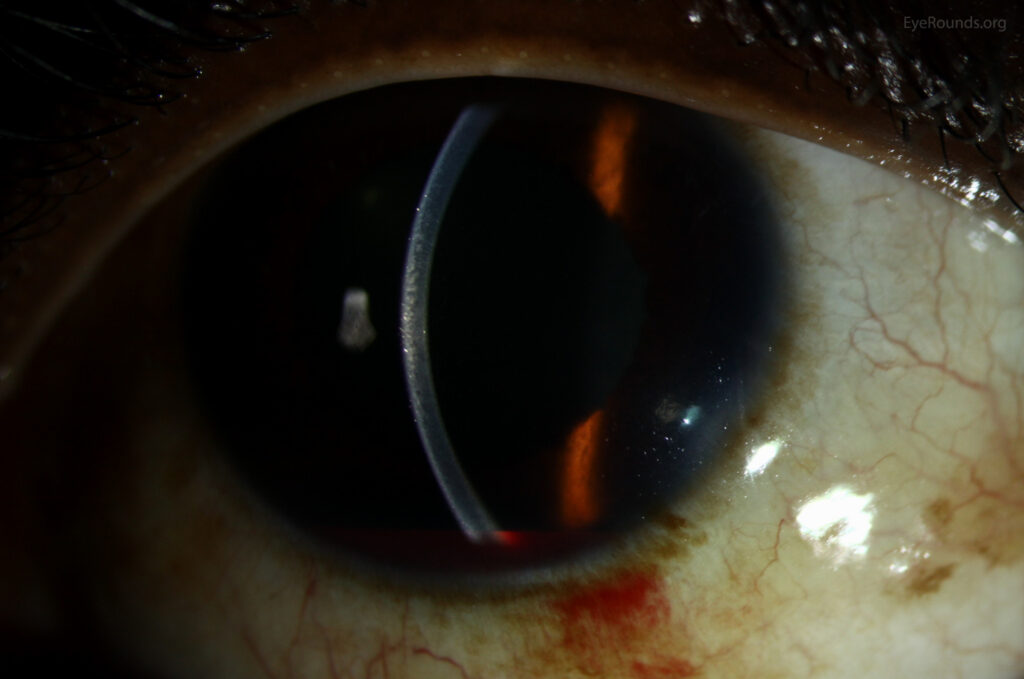
The examiner sees a dark chamber without a visible fluid level.
Iris and pupil details are usually obscured.
Corneal edema may further limit the view when pressure is high.
Therefore, inspect the endothelium and stroma carefully for early blood staining.
Examining the Injured Eye Safely
First, exclude an open globe and protect the eye with a rigid shield.
Measure IOP gently only if the globe appears intact.
Also, avoid scleral depression during the acute phase because it may provoke rebleeding.
Sickle Status in Eight Ball Hyphema
Screen at-risk patients with a sickle solubility test or hemoglobin electrophoresis.
Sickled cells pass poorly through the meshwork, and the optic nerve tolerates raised IOP less well (see sickle cell hyphema).
Therefore, surgical thresholds fall considerably.
Carbonic anhydrase inhibitors and hyperosmotic agents also need caution because they may promote sickling.
Medical Treatment and Its Limits
Initial care includes a shield, head elevation, topical steroid and usually cycloplegia.
Aqueous suppressants then lower IOP, although they cannot clear a clot from the chamber.
Avoid aspirin and other NSAIDs because they may increase bleeding risk.
What the Evidence Shows
A Cochrane review found that antifibrinolytics reduced secondary hemorrhage.
However, it did not show a clear benefit for final visual acuity.
Most trials also included few patients with total hyphema.
Evidence specific to the eight ball presentation therefore remains limited.
When Eight Ball Hyphema Needs Surgery
Commonly cited criteria come from older clinical series, so apply them with judgment.
Consider evacuation for any of these findings:
- Early corneal blood staining at any pressure
- IOP of 50 mmHg or higher for about 4 days
- IOP of 25 mmHg or higher for about 6 days in a total hyphema
- Sickle hemoglobinopathy with IOP of 24 mmHg or higher for over 24 hours
- Pressure that threatens an already damaged optic nerve
Evacuation of Eight Ball Hyphema
Options include paracentesis with irrigation, limbal clot delivery, and removal with a vitrectomy cutter.
The surgeon aims to relieve outflow and pupillary obstruction, and complete clearance is unnecessary.
Avoid pulling adherent clot because this can restart bleeding or injure the iris.
Counseling in Eight Ball Hyphema
Explain that the final visual outcome depends largely on the associated injuries, which may stay hidden initially.
Advise limited activity, strict shield use and urgent return for sudden pain or vision loss.
Also, warn that surgery may become necessary at short notice.
Follow-Up After Eight Ball Hyphema
Review IOP daily during the highest-risk period, because rebleeding commonly occurs within the first several days.
After recovery, perform gonioscopy for angle recession, and look for a cyclodialysis cleft if IOP stays low.
Next, complete a dilated peripheral examination.
Eyes with angle damage need long-term glaucoma surveillance.
Pitfalls to Avoid
- Do not treat a dark total hyphema as routine grade 4
- Never overlook an occult open globe
- Do not delay sickle testing in at-risk patients
- Avoid assuming normal IOP will persist after the first visit


Document what you see
Two smartphone imaging tools built for everyday clinic use — one for the slit lamp, one for the fundus.
From Choroida — the team behind this siteReferences
- Walton W, Von Hagen S, Grigorian R, Zarbin M. Management of traumatic hyphema. Surv Ophthalmol. 2002;47:297-334.
- Gharaibeh A, Savage HI, Scherer RW, Goldberg MF, Lindsley K. Medical interventions for traumatic hyphema. Cochrane Database Syst Rev. 2019;1:CD005431.
- Read J, Goldberg MF. Comparison of medical treatment for traumatic hyphema. Trans Am Acad Ophthalmol Otolaryngol. 1974;78:799-815.
- Brandt MT, Haug RH. Traumatic hyphema: a comprehensive review. J Oral Maxillofac Surg. 2001;59:1462-1470.
- Sankar PS, Chen TC, Grosskreutz CL, Pasquale LR. Traumatic hyphema. Int Ophthalmol Clin. 2002;42:57-68.
Test yourself
A few questions straight from this article.
-
What defines an eight ball hyphema?
Eight ball hyphema is a total hyphema in which dark, purplish-black clot fills the anterior chamber, unlike a bright red, partly mobile total hyphema. -
Why does the blood in an eight ball hyphema turn black?
Impaired aqueous circulation and poor oxygen exchange deoxygenate hemoglobin, so the colour suggests stagnation but does not measure pressure. -
Which slit lamp appearance is typical of an eight ball hyphema?
The examiner sees a dark chamber without a visible fluid level, iris and pupil details are usually obscured, and corneal edema may limit the view. -
Besides trabecular meshwork obstruction, how can clot raise intraocular pressure in eight ball hyphema?
Red cells, fibrin and debris obstruct the meshwork, and clot may also block the pupil and stop aqueous from reaching the angle. -
In a patient with sickle hemoglobinopathy and hyphema, which pressure threshold is cited for considering evacuation?
Sickled cells pass poorly through the meshwork and the optic nerve tolerates raised IOP less well, so evacuation is considered at 24 mmHg or higher for over 24 hours. -
Which pressure-lowering drugs need caution in sickle hemoglobinopathy with hyphema?
Carbonic anhydrase inhibitors and hyperosmotic agents need caution in sickle disease because they may promote sickling. -
Which finding is an indication to consider evacuating an eight ball hyphema at any pressure?
Early corneal blood staining at any pressure is a cited criterion for evacuation, which is why the endothelium and stroma are inspected carefully. -
In traumatic hyphema, what did a Cochrane review find for antifibrinolytics?
Antifibrinolytics reduced secondary hemorrhage but showed no clear benefit for final acuity, and most trials included few total hyphemas. -
During surgical evacuation of an eight ball hyphema, why should adherent clot not be pulled?
Evacuation aims to relieve outflow and pupillary obstruction, complete clearance is unnecessary, and pulling adherent clot can restart bleeding or injure the iris. -
After recovery from an eight ball hyphema, what does gonioscopy look for to guide long-term glaucoma surveillance?
After recovery, gonioscopy looks for angle recession, and eyes with angle damage need long-term glaucoma surveillance.

Writes and reviews the clinical articles on Educate. Every post is checked against peer-reviewed sources before it goes live.