Eyelid skin tag is the common name for an acrochordon, a soft benign outgrowth of eyelid skin.
It is harmless, yet patients often fear cancer when they notice it.
Therefore, confirm the diagnosis and choose a removal method that spares the lid margin.
What an Eyelid Skin Tag Is
An acrochordon is a small fibroepithelial polyp attached to the skin by a narrow stalk.
It usually measures a few millimeters and matches the surrounding skin color.
Malignant change within a true tag is exceptionally rare.
However, several other lesions imitate it closely.
Who Develops Them
Skin tags become more common with age and favor the neck, axillae, groin and eyelids.
They also show an association with obesity, insulin resistance and type 2 diabetes.
As a result, many patients have similar tags on other body sites.
All-fit Slit-Lamp Adapter
Record and share exactly what you see at the slit lamp. One adapter fits any slit lamp or surgical microscope — and any smartphone.
From Choroida — the team behind this siteEyelid Skin Tag at the Slit Lamp
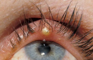
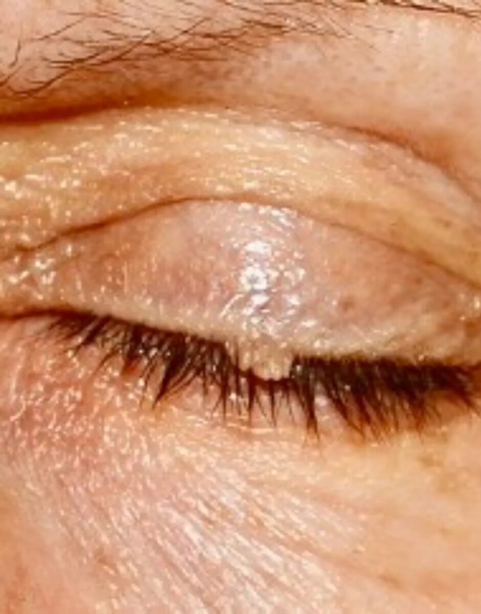
The typical tag is soft, smooth and pedunculated, and it moves freely when touched.
Its surface is slightly wrinkled, without keratin crust or fronds.
The lashes stay intact, and the margin keeps its normal shape.
Next, look for pigment, vessels or ulceration, because these findings change the diagnosis.
What the Patient Reports
Most patients describe a painless soft bump that grew slowly.
Some notice it rubbing on glasses or catching during makeup removal.
Occasionally the tag twists, turns dark and becomes tender after its blood supply fails.
Histology of an Eyelid Skin Tag
Histology shows a loose fibrovascular core covered by thin or mildly acanthotic epidermis.
Therefore, a typical tag lacks the papillary fronds of a squamous papilloma.
The report also confirms the absence of atypia when the clinical picture was uncertain.
Squamous Papilloma Versus Eyelid Skin Tag
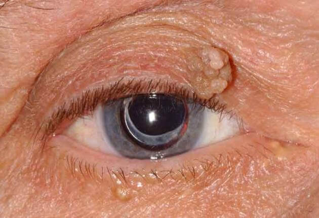
Squamous papilloma is the lesion most often mistaken for a skin tag on the eyelid.
It has a raspberry or finger-like surface, often with keratin and a vessel in each frond.
Still, both lesions are benign, and removal follows similar steps.
Other Benign Look-Alikes
Several benign lesions resemble a tag at first glance (see eyelid lumps).
- Intradermal nevus: dome-shaped, sometimes pigmented, often with hairs
- Seborrheic keratosis: stuck-on, waxy and greasy (see seborrheic keratosis)
- Molluscum contagiosum: pearly papule with central umbilication
- Filiform wart: thin keratotic projections, often near the margin
Red Flags for Malignancy
Basal cell carcinoma can look like a smooth papule, especially on the lower lid.
However, it shows pearly borders, fine telangiectasia or central ulceration.
Lash loss, bleeding, induration and rapid growth also raise suspicion.
In addition, sebaceous carcinoma may appear as margin thickening with madarosis (see sebaceous gland carcinoma).
When to Biopsy an Eyelid Skin Tag
Send the specimen for histopathology whenever any atypical feature exists.
Kersten and colleagues reported that clinical impression alone missed some eyelid malignancies.
Therefore, many oculoplastic surgeons submit every removed lesion.
Recurrence at the same site also requires tissue diagnosis.
Snip Excision
Snip excision suits most pedunculated tags.
First, inject a small volume of local anesthetic, then lift the tag with fine forceps.
Next, cut the stalk flush with the skin using curved scissors.
Pressure or light cautery then controls minor bleeding, and sutures are rarely needed.
Lesions on the Lid Margin
Margin tags demand care to avoid notching and lash loss.
Protect the globe with a corneal shield during the procedure.
Then shave the lesion level with the margin, sparing the tarsus.
Avoid deep cautery near lash follicles, because it can cause trichiasis or madarosis.
Cautery, Radiofrequency and Cryotherapy
Electrocautery and radiofrequency devices remove small tags with little bleeding.
However, thermal artifact may limit histology.
Cryotherapy also treats thin stalks, although it can depigment skin and destroy lash follicles.
As a result, avoid it near the margin and in darker skin.
Home Removal of an Eyelid Skin Tag
Patients often ask about thread ligation, freezing kits or removal creams.
These products target body skin and can injure the lid margin or cornea.
In addition, home removal destroys the specimen and may delay a cancer diagnosis.
Therefore, advise against it for any periocular lesion.
Aftercare and Recurrence
After removal, apply antibiotic ointment for a few days and skip eye makeup.
The site usually heals within one to two weeks.
However, new tags often appear elsewhere, especially with metabolic risk factors.
Counseling for Eyelid Skin Tag Removal
Explain that the lesion is benign and that removal is mainly cosmetic.
Then discuss bleeding, infection, scarring and pigment change.
For margin lesions, also mention possible lash loss or a small notch.
Finally, tell the patient when the histology result will arrive.
Pitfalls to Avoid
- Calling a keratotic, frond-like lesion a simple tag
- Freezing a margin lesion in a patient who values lash appearance
- Discarding tissue from an atypical or recurrent lesion
- Ignoring lash loss, because it often signals malignancy


Document what you see
Two smartphone imaging tools built for everyday clinic use — one for the slit lamp, one for the fundus.
From Choroida — the team behind this siteReferences
- Kersten RC, Ewing-Chow D, Kulwin DR, Gallon M. Accuracy of clinical diagnosis of cutaneous eyelid lesions. Ophthalmology. 1997;104:479-484.
- Deprez M, Uffer S. Clinicopathological features of eyelid skin tumors. A retrospective study of 5504 cases and review of literature. Am J Dermatopathol. 2009;31:256-262.
- Shields JA, Shields CL. Eyelid, Conjunctival, and Orbital Tumors: An Atlas and Textbook. 3rd ed. Philadelphia: Wolters Kluwer; 2016.
- American Academy of Ophthalmology. Basic and Clinical Science Course, Section 7: Oculofacial Plastic and Orbital Surgery. San Francisco: AAO; 2023.
- Bowling B. Kanski’s Clinical Ophthalmology: A Systematic Approach. 8th ed. Edinburgh: Elsevier; 2016.

Writes and reviews the clinical articles on Educate. Every post is checked against peer-reviewed sources before it goes live.