Retinal detachment types fall into three groups: rhegmatogenous, tractional and exudative.
Each type separates the neurosensory retina from the retinal pigment epithelium by a different mechanism.
As a result, the fundus shape, fluid behavior and treatment all differ.
Correct classification at the first visit therefore guides urgency and referral.
Why Retinal Detachment Types Matter
Management follows the mechanism, so a wrong label leads to the wrong treatment.
For example, surgery rarely helps an exudative detachment.
In addition, a missed break in a presumed exudative case delays repair.
Therefore, ask one question first: where does the fluid come from?
Rhegmatogenous Detachment
Rhegmatogenous detachment is the most common type.
A full-thickness retinal break lets liquefied vitreous pass into the subretinal space.
Most breaks follow posterior vitreous detachment because the vitreous pulls at sites of firm adhesion.
Risk factors include myopia, lattice degeneration, cataract surgery and trauma (see rhegmatogenous retinal detachment).
Fundus Explorer Pro
Photograph the retinal findings described here with the phone already in your pocket — 22 D optics and built-in illumination in one handheld unit.
From Choroida — the team behind this siteFundus Signs of Rhegmatogenous Detachment
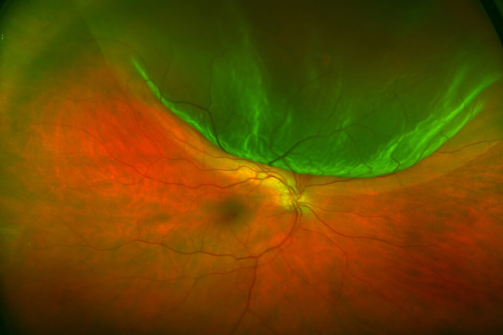
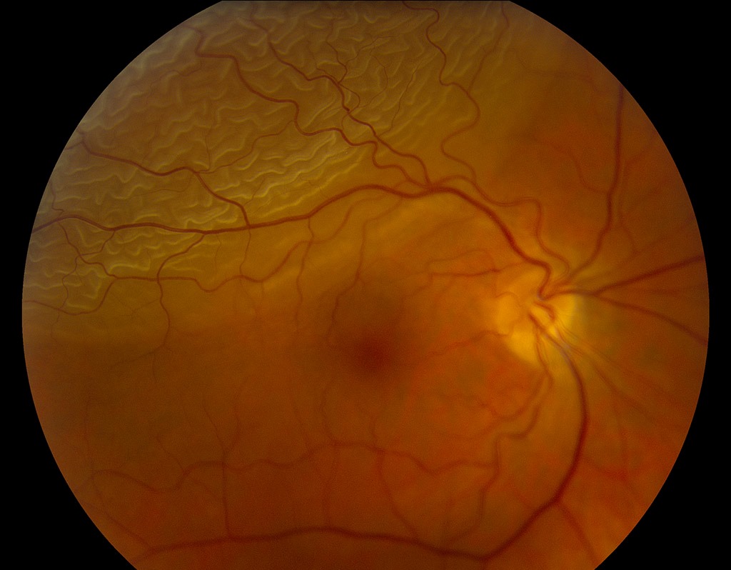
The detached retina looks convex, corrugated and mobile with eye movement.
Pigment cells in the anterior vitreous, called tobacco dust, strongly suggest a break.
Chronic cases also show demarcation lines, thin retina and intraretinal cysts.
Tractional Detachment
Tractional detachment occurs when contracting membranes pull the retina forward without a break.
Proliferative diabetic retinopathy is the classic cause.
Retinopathy of prematurity, sickle cell retinopathy and penetrating trauma also produce it.
Fundus Signs of Tractional Detachment
The retina looks concave, smooth and taut, and it does not move with the eye.
In addition, the detachment follows the vascular arcades and rarely reaches the ora serrata.
On OCT, membranes appear as hyperreflective bands tenting the retina (see tractional retinal detachment).
Exudative Detachment
Exudative, or serous, detachment results from leakage across a damaged blood-retinal barrier.
Neither a break nor traction is present.
Causes include Vogt-Koyanagi-Harada disease, posterior scleritis, choroidal tumors, Coats disease and malignant hypertension.
Preeclampsia and renal failure can also produce it.
Fundus Signs of Exudative Detachment
The retina looks smooth, convex and bullous, usually without folds.
Because the fluid moves freely, it shifts with posture and pools inferiorly.
A choroidal mass or choroidal folds may point to the cause (see exudative retinal detachment).
Shifting Fluid in Retinal Detachment Types
Shifting fluid is the most useful bedside sign of exudative disease.
In contrast, rhegmatogenous fluid moves little because the break anchors its source.
Tractional fluid also stays fixed, while membranes hold the retina in shape.
However, a bullous rhegmatogenous detachment can shift slightly, so always search for a break.
Symptoms Across Retinal Detachment Types
Rhegmatogenous detachment usually starts with flashes, new floaters and a spreading curtain.
Tractional detachment often progresses slowly, with few symptoms until the macula is involved.
Exudative detachment may cause vision that changes with head position.
In addition, inflammatory causes can bring pain, redness or headache.
Mixed Mechanisms in Retinal Detachment Types
Some eyes show more than one mechanism.
For example, contracting diabetic membranes can tear a hole and create a combined detachment.
The retina then becomes more mobile, and the fluid spreads faster.
Therefore, this change usually prompts earlier surgery.
Imaging in Retinal Detachment Types
B-scan ultrasound confirms detachment when media opacity blocks the view.
It also shows tented traction, mobile retina or a choroidal mass.
OCT measures macular involvement, while fluorescein angiography identifies leakage sources in exudative disease.
Treating Rhegmatogenous Detachment
Repair aims to close every break and relieve vitreous traction.
Options include pneumatic retinopexy, scleral buckling and pars plana vitrectomy.
The choice depends on break location, lens status, age and extent.
Then, discuss redetachment risk, cataract and positioning demands with the patient.
Treating Tractional Detachment
Vitrectomy with membrane dissection treats traction that involves or threatens the macula.
However, stable peripheral tractional detachments are often observed.
Preoperative anti-VEGF may reduce bleeding, although delayed surgery risks faster contraction.
Macular ischemia limits the final vision, even after successful reattachment.
Treating Exudative Detachment
Treatment targets the underlying disease.
For example, corticosteroids help Vogt-Koyanagi-Harada disease and posterior scleritis.
Tumors need oncologic care, and Coats disease may need laser to leaking vessels.
However, steroids can worsen central serous chorioretinopathy, so confirm the diagnosis first.
Comparing Retinal Detachment Types
- Rhegmatogenous: break present, convex corrugated retina, little shifting fluid, surgical repair
- Tractional: no break, concave taut retina, fixed fluid, vitrectomy when the macula is threatened
- Exudative: no break, smooth bullous retina, marked shifting fluid, treatment of the cause
Pitfalls to Avoid
- Do not call a detachment exudative before scleral depression excludes a break
- Never start steroids for serous fluid without excluding central serous chorioretinopathy
- Remember that a new break can convert a tractional detachment into a combined one


Document what you see
Two smartphone imaging tools built for everyday clinic use — one for the slit lamp, one for the fundus.
From Choroida — the team behind this siteReferences
- Ghazi NG, Green WR. Pathology and pathogenesis of retinal detachment. Eye (Lond). 2002;16:411-421.
- Mitry D, Fleck BW, Wright AF, et al. Pathogenesis of rhegmatogenous retinal detachment: predisposing anatomy and cell biology. Retina. 2010;30:1561-1572.
- Amer R, Nalcı H, Yalçındağ N. Exudative retinal detachment. Surv Ophthalmol. 2017;62:723-769.
- Flaxel CJ, Adelman RA, Bailey ST, et al. Posterior vitreous detachment, retinal breaks, and lattice degeneration Preferred Practice Pattern. Ophthalmology. 2020;127:P146-P181.

Writes and reviews the clinical articles on Educate. Every post is checked against peer-reviewed sources before it goes live.