Drusen vs pseudodrusen is a common comparison in age-related macular degeneration (AMD) clinics.
Both appear as yellowish deposits in the posterior pole of older adults.
However, they sit on opposite sides of the retinal pigment epithelium (RPE).
As a result, they differ in imaging, visual function and progression risk.
Anatomy in Drusen vs Pseudodrusen
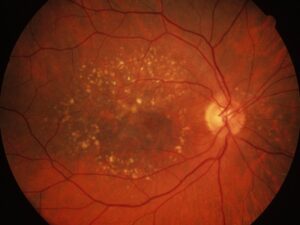
Soft drusen collect beneath the RPE, inside Bruch’s membrane.
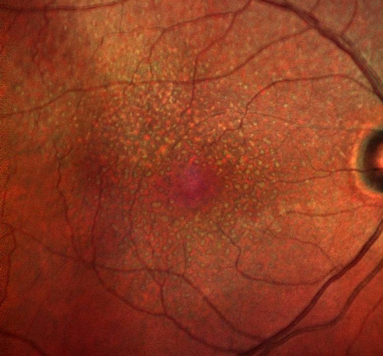
Reticular pseudodrusen lie above the RPE, under the photoreceptor outer segments.
For this reason, many authors call them subretinal drusenoid deposits (SDD).
Zweifel and colleagues also confirmed this location with histology and OCT.
Fundus Color in Drusen vs Pseudodrusen
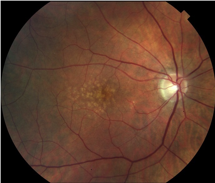
Soft drusen look yellow-white with blurred edges and cluster in the central macula.
In contrast, pseudodrusen look paler, sometimes bluish, and form an interlacing network.
They favor the superior macula and often extend beyond the arcades.
Also, they can be faint on color photos.
Fundus Explorer Pro
Photograph the retinal findings described here with the phone already in your pocket — 22 D optics and built-in illumination in one handheld unit.
From Choroida — the team behind this siteExamination Tips
First, use a high-power lens with good stereopsis.
Soft drusen show clear elevation, while pseudodrusen look flat and poorly defined.
Blue or red-free light also improves pseudodrusen contrast.
Therefore, a normal clinical view does not exclude SDD.
Multimodal Imaging in Drusen vs Pseudodrusen
No single test separates the two in every eye.
Instead, combine near-infrared reflectance, autofluorescence and OCT.
Near-infrared reflectance then shows pseudodrusen as dark dots, often with a bright halo.
Autofluorescence Findings
Soft drusen are usually isoautofluorescent or mildly hyperautofluorescent.
However, their signal varies with the health of the overlying RPE.
By contrast, pseudodrusen produce a reticular pattern of small hypoautofluorescent spots.
For signal interpretation, see fundus autofluorescence imaging.
OCT in Drusen vs Pseudodrusen
OCT gives the most direct answer.
Soft drusen lift the RPE band into smooth domes with medium reflectivity.
Meanwhile, Bruch’s membrane stays flat beneath them.
In addition, pseudodrusen form granular or conical deposits above the RPE band.
Staging Subretinal Deposits
Zweifel described four OCT stages of SDD.
First, granular material sits between the RPE and ellipsoid zone.
Next, mounds deform the ellipsoid zone, then conical lesions break through it.
Finally, deposits may regress and leave outer retinal thinning.
Choroid and Function
Eyes with pseudodrusen also often show a thin choroid on OCT.
Patients may report poor night vision and slow adaptation to dim light.
However, visual acuity can remain normal despite these symptoms.
AMD Risk in Drusen vs Pseudodrusen
Large soft drusen define intermediate AMD in the Beckman classification.
Medium drusen measure 63 to 125 µm, while large drusen exceed 125 µm.
Pigmentary abnormalities also add risk.
In addition, pseudodrusen carry independent risk (see soft drusen versus hard drusen).
Patterns of Progression
Soft drusen classically progress toward geographic atrophy or type 1 neovascularization.
In contrast, pseudodrusen link more often with geographic atrophy and type 3 neovascularization.
Because many eyes have both, risks overlap.
When Both Coexist
Mixed phenotypes are common in advanced age.
In such eyes, grade drusen size first, then record pseudodrusen extent.
Also, note the quadrants involved, because widespread deposits may signal higher risk.
Differential Diagnosis
Several lesions mimic either deposit (see drusen vs exudates):
- Hard drusen and cuticular drusen
- Hard exudates in vascular disease
- Pattern dystrophy and vitelliform lesions
- Calcified or regressing drusen
Detecting Pseudodrusen in Routine Practice
First, order near-infrared reflectance when a patient reports poor dark adaptation.
Next, scan the superior macula and the area beyond the arcades on OCT.
Then confirm the finding on at least two modalities.
Because SDD are easy to miss, a systematic review prevents under-grading.
Management of Drusen vs Pseudodrusen
No treatment safely removes either deposit.
However, the AREDS formulation reduced progression in intermediate AMD, and AREDS2 refined it.
Yet AREDS grading used color photos, which often miss pseudodrusen.
Then add smoking cessation and home Amsler monitoring.
Laser and Pseudodrusen
The LEAD trial tested subthreshold nanosecond laser in intermediate AMD.
Overall, it did not slow progression to late AMD.
However, a post hoc analysis suggested possible harm in eyes with pseudodrusen.
As a result, avoid laser for drusen outside trials.
Counseling Patients with Drusen
First, explain that neither deposit means certain vision loss.
However, pseudodrusen suggest a higher chance of atrophy, so regular review matters.
Also, advise a healthy diet and no smoking.
Follow-up in Drusen vs Pseudodrusen
Many clinicians review intermediate AMD every six to twelve months.
Next, repeat OCT and near-infrared imaging at each visit.
Finally, teach patients to report new distortion quickly.
Pitfalls to Avoid
- However, do not call flat pseudodrusen early drusen on color photos alone
- Therefore, add near-infrared imaging when SDD are suspected
- Also check the area above the superior arcade
- Then grade risk from both deposit types, not drusen size alone


Document what you see
Two smartphone imaging tools built for everyday clinic use — one for the slit lamp, one for the fundus.
From Choroida — the team behind this siteReferences
- Zweifel SA, Spaide RF, Curcio CA, et al. Reticular pseudodrusen are subretinal drusenoid deposits. Ophthalmology. 2010;117:303-312.
- Spaide RF, Ooto S, Curcio CA. Subretinal drusenoid deposits AKA pseudodrusen. Surv Ophthalmol. 2018;63:782-815.
- Ferris FL, Wilkinson CP, Bird A, et al. Clinical classification of age-related macular degeneration. Ophthalmology. 2013;120:844-851.
- Age-Related Eye Disease Study 2 Research Group. Lutein + zeaxanthin and omega-3 fatty acids for age-related macular degeneration: the AREDS2 randomized clinical trial. JAMA. 2013;309:2005-2015.
- Guymer RH, Wu Z, Hodgson LAB, et al. Subthreshold nanosecond laser intervention in age-related macular degeneration: the LEAD randomized controlled clinical trial. Ophthalmology. 2019;126:829-838.

Writes and reviews the clinical articles on Educate. Every post is checked against peer-reviewed sources before it goes live.