
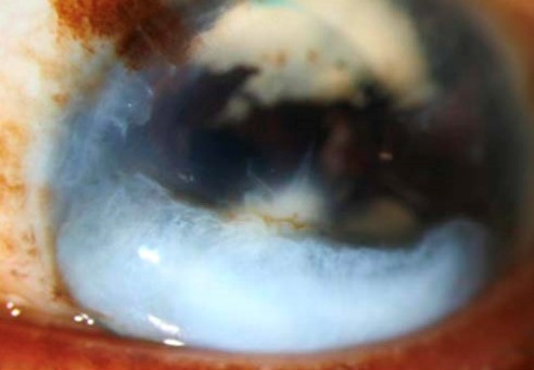
A corneal keloid is a genuinely rare entity, a dense, elevated, fibrous overgrowth of corneal tissue that, while sharing its name and some histologic similarity with cutaneous keloids, follows its own distinct clinical pattern and is far less common than its skin counterpart, often developing after significant corneal injury, inflammation, or surgery, particularly in younger patients.
What Makes This Different From an Ordinary Corneal Scar
Unlike an ordinary corneal scar, which represents a relatively organized fibrous healing response proportionate to the original injury, a corneal keloid involves an excessive, disorganized fibrous tissue proliferation extending beyond what would be expected from the inciting injury or inflammatory process alone, producing a distinctly elevated, often quite firm and white mass rather than a flatter, more typical corneal opacity.
Clinical Features
- A white to grayish-white, densely fibrous, elevated mass on the corneal surface, often with a firm, somewhat irregular texture distinguishing it from smoother corneal opacities
- A history of prior significant corneal injury, inflammation, or surgery in the affected eye is typical, though the interval between the inciting event and keloid development can be considerable
- More frequently reported in children and younger patients, though it can occur across a range of ages
- Vision is often significantly affected when the lesion involves the central cornea, given both its opacity and its irregular, elevated surface contour
All-fit Slit-Lamp Adapter
Record and share exactly what you see at the slit lamp. One adapter fits any slit lamp or surgical microscope — and any smartphone.
From Choroida — the team behind this siteDiagnostic Evaluation
- Slit-lamp examination characterizing the lesion’s appearance, size, and relationship to the visual axis
- A detailed history of any prior corneal injury, inflammation, infection, or surgery in the affected eye, relevant to understanding the likely inciting event
- Differentiation from other causes of corneal opacity and elevation, including other reparative or proliferative corneal conditions, based on the characteristic clinical appearance and history
- Histopathologic examination, when tissue is obtained through excision, can confirm the diagnosis based on characteristic dense, disorganized collagen proliferation
Management
Observation for Stable, Visually Insignificant Lesions
Small, peripheral, or visually insignificant corneal keloids can be observed, given the generally stable, though sometimes very slowly progressive, natural history these lesions can follow.
Surgical Excision
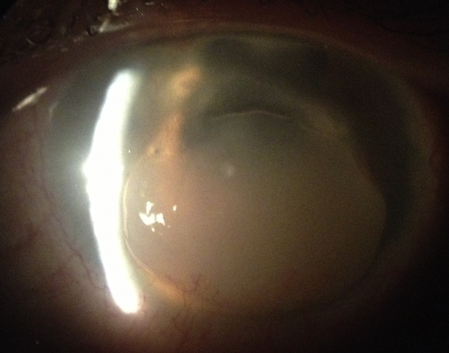
Surgical excision is considered for visually significant lesions, particularly those involving the central visual axis, aiming to restore corneal clarity and improve vision, with the surgical approach, ranging from superficial keratectomy to, for deeper or more extensive lesions, lamellar or penetrating keratoplasty, tailored to the depth and extent of the individual lesion.
The Real Risk of Recurrence
An important management consideration specific to this condition is a genuine tendency toward recurrence after excision, reflecting the same excessive, disorganized fibrous proliferative tendency responsible for the original lesion, and this recurrence risk should be part of pre-surgical counseling, along with close postoperative monitoring to allow early identification and management of any recurrent proliferation.
Differential diagnosis
A corneal keloid should be distinguished from a dense scar, which is not elevated and does not grow, from a limbal dermoid, which is congenital, from Salzmann nodular degeneration, which is bluish and nodular, and from a fibrous plaque of a neoplasm such as ocular surface squamous neoplasia. The history of injury or inflammation and the growth over time support the diagnosis. Histology shows thick, disorganized collagen with fibroblasts, and no inflammation in the later stage.
Surgical considerations
The surgeon chooses between superficial keratectomy and lamellar or penetrating keratoplasty, depending on the depth. Adjunctive measures such as steroid drops and, in some centers, antimetabolites have been used to reduce recurrence, although the evidence is limited and patients should be told that recurrence can occur. Because the lesion is rare, referral to a corneal specialist is sensible.
Follow-up
Review at short intervals in the first months, then at longer intervals. Take photographs of the cornea and keep records of the visual acuity so that early recurrence can be recognized. Counsel patients to report changes in vision, redness, or a new white area on the cornea.
Counselling about expectations
Explain that the lesion is benign but can be stubborn, that surgery may need to be repeated, and that glasses or contact lenses may be needed afterward to correct the vision. Patients with a history of keloids elsewhere should mention this, since a tendency to excessive scarring is relevant to surgical planning.
Prognosis
Corneal keloid, once surgically excised, carries a meaningfully higher recurrence risk than more typical corneal scarring or opacity, reflecting its underlying excessive fibroproliferative tendency, and this recurrence potential should shape both surgical planning and the ongoing follow-up plan after excision.
Individualized management, weighing the degree of visual impact against the genuine risk of recurrent proliferation after surgical intervention, supports the most appropriate approach for a given patient, with careful, sustained follow-up remaining important regardless of which specific management path is chosen.


Document what you see
Two smartphone imaging tools built for everyday clinic use — one for the slit lamp, one for the fundus.
From Choroida — the team behind this siteReferences
- Lahoti S, Weiss M. Corneal keloid. Am J Ophthalmol. 1989;108:305-306.
- Bourgeois J, Yau B, Wilson DJ, Selva D. Corneal keloid. Cornea. 2011;30:1493-1495.
- Rao SK, Fogla R, Rajagopal R, Sitalakshmi G, Padmanabhan P. Corneal keloid: report of two cases. Indian J Ophthalmol. 1999;47:181-183.
- Farrell TA, Alward WL, Verdick RE, Nerad JA. Corneal keloid. Am J Ophthalmol. 1993;116:247-249.

Writes and reviews the clinical articles on Educate. Every post is checked against peer-reviewed sources before it goes live.


