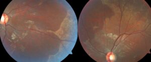
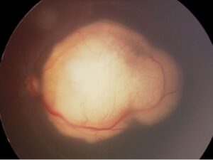
Vitreous hemorrhage recovery depends on the bleeding source, the density of blood and the state of the gel.
Mild bleeding may clear within weeks, while dense hemorrhage can persist for months.
Therefore, clinicians should offer a time range after establishing the cause (see the diagnostic approach).
How Blood Clears From the Vitreous
Red cells lyse inside the gel, and macrophages then remove the debris.
However, the vitreous clears blood slowly because it has no vessels of its own.
As a result, vision improves in gradual steps.
Vitreous Hemorrhage Recovery Timeline
However, no study provides a reliable clock, so these estimates are rough:
- First days: blood settles inferiorly, so the superior fundus may appear first
- Two to six weeks: light hemorrhage often thins and acuity may improve
- Two to three months: moderate hemorrhage may still be clearing
- Beyond three months: dense or recurrent blood often prompts a surgical discussion
Fundus Explorer Pro
Photograph the retinal findings described here with the phone already in your pocket — 22 D optics and built-in illumination in one handheld unit.
From Choroida — the team behind this siteWhat Patients Notice During Vitreous Hemorrhage Recovery
Patients describe a red haze, dark floaters or cobweb shadows.
Vision may seem worse after waking because blood redistributes during sleep.
A sudden new shower, however, suggests fresh bleeding and needs prompt review.
What Slows Vitreous Hemorrhage Recovery
Several factors extend clearing time:
- Dense blood that hides the fundus
- Recurrent bleeding from active neovascularization
- Fibrovascular traction that keeps tearing vessels
- Formed gel, which holds blood longer than liquefied vitreous
How the Source Shapes the Forecast
A posterior vitreous detachment can cause a single bleed, although a retinal tear must be excluded.
In contrast, proliferative diabetic retinopathy can bleed repeatedly until the neovascular drive is controlled.
Therefore, the forecast improves only after the source is treated.
Monitoring Vitreous Hemorrhage Recovery With No Fundus View
Observation is safe only when the clinician can monitor the retina.
Therefore, B-scan ultrasonography should accompany each visit when blood blocks the view.
It can reveal a retinal detachment, a flap tear or a mass.
A normal scan does not exclude a small tear, so repeat examination remains necessary.
In addition, check the iris and angle for neovascularization.
Activity, Position and Antithrombotic Drugs
Many clinicians advise raising the head of the bed so gravity moves blood from the visual axis.
Evidence for this advice is limited, so present it as supportive.
Aspirin or anticoagulants may increase bleeding, but patients often take them for stroke prevention.
Therefore, discuss any change with the prescribing physician.
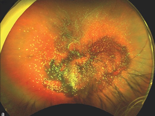
Complications That Delay Vitreous Hemorrhage Recovery
Retinal detachment is the most urgent complication.
Ghost cell glaucoma can also develop when degenerated red cells block the trabecular meshwork.
Neovascular glaucoma can follow when dense blood delays laser treatment in an ischemic eye.
Diabetic Eyes: Injection or Surgery?
Protocol N compared ranibizumab with saline and found no significant difference in vitrectomy rates by 16 weeks.
Protocol AB then compared aflibercept with vitrectomy plus panretinal photocoagulation (see the Protocol AB review).
Mean acuity over 24 weeks did not differ significantly, although vitrectomy improved vision faster.
However, some aflibercept eyes later needed surgery.
The Diabetic Retinopathy Vitrectomy Study favored early surgery in severe type 1 diabetic hemorrhage, but it predates modern anti-VEGF therapy.
When Observation Should Give Way to Vitrectomy
No single deadline fits every patient.
Consider surgery when any of these apply:
- Ultrasound shows retinal detachment or a tear
- Traction threatens the macula
- Dense hemorrhage shows no clearing after several weeks to months
- Neovascularization develops without a laser view
- Ghost cell glaucoma resists medical treatment
Vitreous Hemorrhage Recovery After Vitrectomy
Pars plana vitrectomy removes the blood and allows immediate treatment of the source.
Vision can therefore improve within days to weeks.
However, gas tamponade delays useful vision until the bubble absorbs.
Surgery cannot restore function lost to macular ischemia.
Visual Prognosis Depends on the Macula
Final acuity reflects retinal health once the media clear.
For example, an uncomplicated bleed after posterior vitreous detachment often carries a favorable outlook.
In contrast, macular ischemia or detachment may limit recovery.
Counseling on Vitreous Hemorrhage Recovery
First, explain that clearing is slow and progress may fluctuate.
Next, give warning symptoms, including new floaters, flashes, a curtain or pain.
Finally, avoid promising a date, because rebleeding can reset the timeline.
Pitfalls to Avoid
- Observing a dense hemorrhage without ultrasound
- Assuming a diabetic bleed without excluding a tear
- Equating clearing with cure while neovascularization remains active


Document what you see
Two smartphone imaging tools built for everyday clinic use — one for the slit lamp, one for the fundus.
From Choroida — the team behind this siteReferences
- Spraul CW, Grossniklaus HE. Vitreous hemorrhage. Surv Ophthalmol. 1997;42:3-39.
- Sarrafizadeh R, Hassan TS, Ruby AJ, et al. Incidence of retinal detachment and visual outcome in eyes presenting with posterior vitreous separation and dense fundus-obscuring vitreous hemorrhage. Ophthalmology. 2001;108:2273-2278.
- Diabetic Retinopathy Clinical Research Network. Randomized clinical trial evaluating intravitreal ranibizumab or saline for vitreous hemorrhage from proliferative diabetic retinopathy. JAMA Ophthalmol. 2013;131:283-293.
- Diabetic Retinopathy Vitrectomy Study Research Group. Early vitrectomy for severe vitreous hemorrhage in diabetic retinopathy: two-year results. Arch Ophthalmol. 1985;103:1644-1652.

Writes and reviews the clinical articles on Educate. Every post is checked against peer-reviewed sources before it goes live.