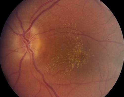
Macular star is a radial pattern of hard exudates centered on the fovea.
It reflects lipid leakage, usually from a swollen optic disc or nearby vessels.
Therefore, the sign points the examiner toward the disc, the blood pressure, or an infection.
What a Macular Star Looks Like
Yellow-white exudates line up like spokes around the foveal center.
The pattern may be complete or may cover only part of the macula.
A partial star on the nasal side is common, because the leakage starts at the disc.
In addition, the foveal center often stays relatively clear early on.
Why Exudates Form a Star
The anatomy of Henle fiber layer explains the shape.
Near the fovea, photoreceptor axons run obliquely and radially away from the center.
Lipid and protein that leak into the outer plexiform layer settle between these fibers.
As a result, the deposits follow the fiber direction and form spokes.
Fundus Explorer Pro
Photograph the retinal findings described here with the phone already in your pocket — 22 D optics and built-in illumination in one handheld unit.
From Choroida — the team behind this siteWhere the Fluid Comes From
In most cases, the macular vessels are not the source.
Instead, incompetent capillaries at the swollen disc leak serous fluid.
The fluid tracks within and under the retina toward the macula.
Then the water resorbs, while the lipid stays behind and precipitates.
Timing of Macular Star and Disc Edema
Disc swelling usually comes first.
The star often appears one to two weeks later, so a first visit may show disc edema alone.
Therefore, repeat the fundus examination when you suspect neuroretinitis.
Disc edema usually settles over several weeks, while the star may persist for six to twelve months.
Neuroretinitis and the Macular Star
Neuroretinitis is the classic cause in young, healthy patients (see neuroretinitis).
Patients typically report painless, unilateral blurred vision.
Bartonella henselae, the agent of cat scratch disease, is the most frequently identified cause.
Other infections include syphilis, Lyme disease, toxoplasmosis and tuberculosis.
When testing finds no cause, the label idiopathic stellate neuroretinitis applies.
Hypertension and Star Exudates
Severe hypertension can produce a star together with disc swelling.
This picture belongs to malignant or grade 4 hypertensive retinopathy.
Flame hemorrhages, cotton wool spots and arteriolar narrowing usually accompany it.
In addition, the findings are often bilateral, which separates them from neuroretinitis.
Macular Star in Papilledema
Chronic or severe papilledema occasionally produces a partial star.
The exudates usually sit nasally, between the disc and the fovea.
However, papilledema is bilateral, and the patient may report headache or transient obscurations.
Workup then follows the usual papilledema pathway (see papilledema).
Other Causes of a Macular Star
Several other conditions can leak lipid into Henle fiber layer.
- Retinal vein occlusion with disc edema
- Diabetic papillopathy
- Anterior ischemic optic neuropathy (uncommon)
- Retinal arterial macroaneurysm near the macula
- Toxocariasis and other posterior segment infections
OCT Findings in Macular Star
OCT shows hyperreflective deposits in the outer plexiform layer.
In neuroretinitis, peripapillary subretinal fluid often extends toward the fovea.
Also, serial scans help you follow resorption of the fluid.
Fluorescein Angiography
Angiography in neuroretinitis shows leakage from the disc vessels.
By contrast, the perifoveal capillaries usually do not leak.
This pattern helps separate neuroretinitis from primary macular disease (see macular edema causes).
Workup for a Macular Star
First, measure blood pressure in the clinic.
Next, take an exposure history covering cats, ticks, travel and sexual risk.
Then order Bartonella and syphilis serology, adding other tests as the history suggests.
Bilateral disc swelling calls for neuroimaging before you attribute it to infection.
Visual Symptoms and Prognosis
Vision may fall significantly at onset, often with a central or cecocentral scotoma.
A relative afferent pupillary defect may be present, although it is often mild.
Most patients with neuroretinitis regain good vision over months.
However, some keep subtle color or contrast deficits.
Treatment of Macular Star Causes
Treatment targets the cause, since the exudates resolve slowly once leakage stops.
For Bartonella neuroretinitis, clinicians often prescribe doxycycline, sometimes with rifampin.
However, controlled trial evidence for antibiotics is lacking, and many cases recover untreated.
Some clinicians add systemic corticosteroids, although their benefit remains uncertain.
Managing Hypertensive Macular Star
Hypertensive emergency needs medical care alongside the eye examination.
Blood pressure should come down in a controlled way, under physician supervision.
Then the disc swelling and exudates fade gradually over months.
Counseling the Patient
Explain that the yellow spots are lipid deposits left behind by the leak.
Also warn that they may look worse for a few weeks before they clear.
Patients accept slow recovery better when they know the expected timeline.
Pitfalls to Avoid
- Do not dismiss isolated disc edema because no star is present yet
- Never skip blood pressure measurement
- Avoid labeling bilateral disc swelling as neuroretinitis without imaging
- Do not assume the macular vessels leak without checking angiography
For the latest studies, see the PubMed results for macular star.


Document what you see
Two smartphone imaging tools built for everyday clinic use — one for the slit lamp, one for the fundus.
From Choroida — the team behind this siteReferences
- Dreyer RF, Hopen G, Gass JD, Smith JL. Leber’s idiopathic stellate neuroretinitis. Arch Ophthalmol. 1984;102:1140-1145.
- Purvin V, Sundaram S, Kawasaki A. Neuroretinitis: review of the literature and new observations. J Neuroophthalmol. 2011;31:58-68.
- Suhler EB, Lauer AK, Rosenbaum JT. Prevalence of serologic evidence of cat scratch disease in patients with neuroretinitis. Ophthalmology. 2000;107:871-876.
- Wong TY, Mitchell P. Hypertensive retinopathy. N Engl J Med. 2004;351:2310-2317.
- Gass JDM. Stereoscopic Atlas of Macular Diseases: Diagnosis and Treatment. 4th ed. St Louis: Mosby; 1997.

Writes and reviews the clinical articles on Educate. Every post is checked against peer-reviewed sources before it goes live.