Pingueculitis is acute inflammation of a pre-existing pinguecula on the bulbar conjunctiva.
The yellow nodule becomes red, raised and irritable within days.
Patients often worry about infection or a growth.
However, a careful slit-lamp examination and one simple test usually settle the diagnosis.
What Pingueculitis Is
A pinguecula is a degenerative deposit of altered collagen in the interpalpebral conjunctiva.
It usually sits next to the nasal limbus.
Pingueculitis occurs when the lesion and the surrounding conjunctiva become inflamed (see pinguecula basics).
Why the Lesion Becomes Inflamed
The raised deposit disturbs the local tear film.
As a result, the surface over it dries between blinks.
Then exposure and microtrauma provoke a local inflammatory response.
However, the exact mechanism is not fully defined, although tear film instability appears to contribute.
All-fit Slit-Lamp Adapter
Record and share exactly what you see at the slit lamp. One adapter fits any slit lamp or surgical microscope — and any smartphone.
From Choroida — the team behind this siteCommon Triggers of Pingueculitis
Ultraviolet light, wind, dust and dry air are the usual triggers.
Air conditioning and long screen sessions also reduce blinking and dry the surface.
In addition, contact lens edges can rub a raised lesion.
Also, coexisting dry eye or blepharitis makes flares more frequent.
Symptoms Patients Report
Most patients notice a red patch at the side of one eye.
They also describe grittiness, burning or a foreign body sensation.
However, pain is usually mild and superficial.
Vision stays normal, because the lesion lies outside the visual axis.
Slit-Lamp Signs in Pingueculitis
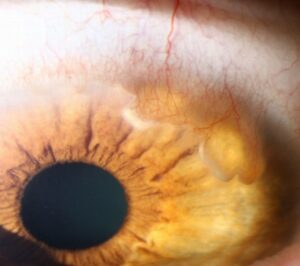
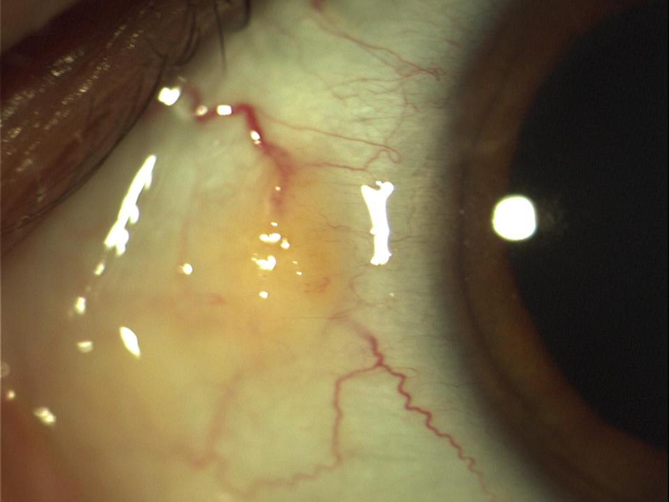
First, the examiner sees a yellow-white, slightly elevated nodule near the limbus.
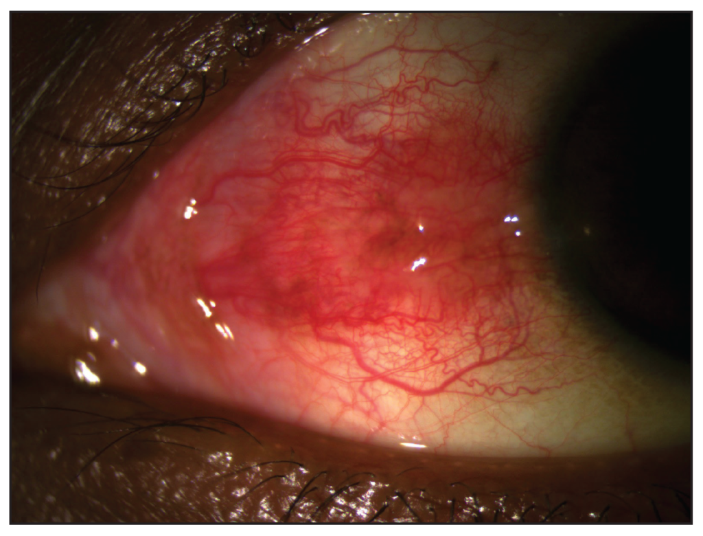
Dilated conjunctival vessels surround and cross the lesion.
Fluorescein may also show punctate staining over its surface.
However, when elevation is marked, the adjacent cornea may develop a small dellen.
Separating Pingueculitis From Episcleritis
Episcleritis causes sectoral or nodular redness of episcleral vessels, often without a yellow deposit.
In pingueculitis, however, the redness centers on a visible pinguecula.
Conjunctival vessels also slide freely when you push them with a cotton-tipped applicator (see episcleritis).
When to Suspect Nodular Scleritis
Nodular scleritis produces deep, boring pain that may wake the patient.
The nodule is immobile, tender and violaceous in daylight.
Furthermore, the deep vascular plexus stays engorged after phenylephrine.
Therefore, any of these features requires a full workup (see scleritis).
The Phenylephrine Test
Topical phenylephrine 10% blanches conjunctival and superficial episcleral vessels within minutes.
In pingueculitis, the redness therefore fades almost completely.
However, persistent deep redness suggests scleral involvement.
Next, check the eye in daylight for a bluish hue.
Other Mimics to Exclude
- Inflamed pterygium with corneal invasion
- Ocular surface squamous neoplasia with leukoplakia or feeder vessels
- Also, a phlyctenule at the apex of a vascular wedge
- Retained foreign body or granuloma
- Finally, conjunctival lymphoma with salmon-pink patches
Treatment Principles for Pingueculitis
The goal is to calm inflammation and protect the surface.
Most flares settle within days to a few weeks with conservative care.
Therefore, start with preservative-free lubricants, cold compresses and trigger control.
Then reserve anti-inflammatory drops for persistent or bothersome cases.
Short Steroid Courses
A mild topical steroid such as fluorometholone or loteprednol speeds resolution.
Therefore, use a short course, typically one to two weeks with a taper.
Also check intraocular pressure if treatment runs longer.
Steroids calm the redness, while the pinguecula itself stays unchanged.
Topical NSAIDs as an Alternative
Topical NSAIDs avoid steroid-related pressure rise.
A randomized trial reported comparable improvement with indomethacin and dexamethasone for inflamed pinguecula and pterygium.
However, NSAIDs can sting and, rarely, cause corneal melting with prolonged use.
Therefore, keep courses brief and monitor the epithelium.
Prevention of Recurrent Pingueculitis
Wraparound sunglasses reduce ultraviolet and wind exposure outdoors.
Regular lubrication also helps patients with coexisting dry eye.
In addition, treat blepharitis and refit contact lenses that rub the lesion.
When Surgery Is Considered
Excision is uncommon for this condition.
Surgeons consider it for frequent recurrences, contact lens intolerance or cosmetic concern.
Before referral, discuss scarring and the chance of regrowth.
Counseling Patients With Pingueculitis
First, explain that a typical lesion is benign.
Then tell patients that the yellow spot will remain after the redness clears.
Also advise urgent review for severe pain, photophobia or reduced vision.
As a result, patients who recognize their triggers usually manage future flares early.
Pitfalls to Avoid
- Do not label every nodular red eye as an inflamed pinguecula without the phenylephrine test
- Never continue topical steroids for weeks without checking pressure
- Also watch for neoplasia in gelatinous, leukoplakic or atypical lesions
- Avoid antibiotic drops for a simple sterile flare
For the latest studies, see the PubMed results for pingueculitis.


Document what you see
Two smartphone imaging tools built for everyday clinic use — one for the slit lamp, one for the fundus.
From Choroida — the team behind this siteReferences
- Watson PG, Hayreh SS. Scleritis and episcleritis. Br J Ophthalmol. 1976;60:163-191.
- Frucht-Pery J, Siganos CS, Solomon A, et al. Topical indomethacin solution versus dexamethasone solution for treatment of inflamed pterygium and pinguecula: a prospective randomized clinical study. Am J Ophthalmol. 1999;127:148-152.
- Dong N, Li W, Lin H, et al. Abnormal epithelial differentiation and tear film alteration in pinguecula. Invest Ophthalmol Vis Sci. 2009;50:2710-2715.
- Bowling B. Kanski’s Clinical Ophthalmology: A Systematic Approach. 8th ed. Elsevier; 2016.
- American Academy of Ophthalmology. Basic and Clinical Science Course, Section 8: External Disease and Cornea. San Francisco: AAO; 2023.

Writes and reviews the clinical articles on Educate. Every post is checked against peer-reviewed sources before it goes live.