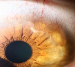
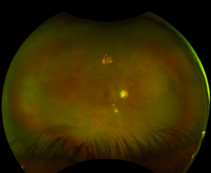
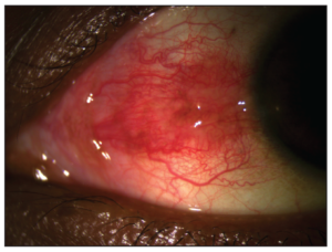
Iridodonesis is a visible tremor of the iris that appears with eye movement.
It occurs when the iris loses its normal support from the lens behind it.
Therefore, the sign points the examiner toward the zonules, the lens and the capsule.
A short look at the slit lamp often answers a surgical question early.
What Iridodonesis Is
The posterior iris normally rests on the anterior lens surface near the pupil.
That contact damps iris movement during saccades.
When the lens shifts backward, tilts or disappears, the iris loses this backstop.
As a result, the iris diaphragm ripples briefly after each eye movement.
Why the Iris Shakes
The aqueous moves with inertia when the globe rotates and then stops.
Because nothing firm supports the pupillary zone, the iris follows that fluid wave.
The tremor is usually most visible near the pupil margin.
A deep, uneven anterior chamber often accompanies it.
All-fit Slit-Lamp Adapter
Record and share exactly what you see at the slit lamp. One adapter fits any slit lamp or surgical microscope — and any smartphone.
From Choroida — the team behind this siteIridodonesis and Phacodonesis
Phacodonesis describes movement of the lens itself.
Iridodonesis describes movement of the iris, which is thinner and easier to see.
However, the two signs frequently coexist in eyes with zonular loss.
In addition, aphakic and some pseudophakic eyes show iris tremor without any natural lens.
Causes of Iridodonesis
- Aphakia after intracapsular or complicated cataract surgery
- Pseudoexfoliation syndrome with weak zonules
- Blunt trauma with zonular dialysis
- Marfan syndrome and other causes of ectopia lentis
- Homocystinuria and Weill-Marchesani syndrome
- High myopia and hypermature cataract
- Sulcus or anterior chamber IOLs, or a loose capsular bag
Eliciting Iridodonesis at the Slit Lamp
First, examine the patient before dilation, because a dilated pupil hides the sign.
Use a moderately wide beam at low to medium magnification.
Next, ask the patient to look quickly to one side and then straight ahead.
Then watch the iris for a few seconds after the eye stops.
Also compare both eyes, since asymmetry is more telling than a faint tremor in one eye.
Other Signs That Support the Finding
- Asymmetric anterior chamber depth between eyes or quadrants
- Visible lens equator or zonules through the dilated pupil
- Pseudoexfoliation material on the pupil margin or capsule
- Vitreous strands in the anterior chamber
- Pupillary block from a forward-shifted lens (see iris bombe)
Iridodonesis in Pseudoexfoliation
Pseudoexfoliation is a common acquired cause of zonular weakness in older adults.
Fibrillar material deposits on the zonules and weakens their attachments.
As a result, iris tremor in these eyes predicts fragile support during phacoemulsification.
In addition, these pupils often dilate poorly, which adds surgical difficulty.
Iridodonesis After Trauma
Blunt injury can tear zonules in one sector.
Therefore, look for localized tremor, a deeper chamber in one quadrant and iridodialysis.
Angle recession and hyphema often accompany the zonular damage.
Also check for a cyclodialysis cleft when the pressure is unexpectedly low (see cyclodialysis cleft).
Iridodonesis in Aphakia and Pseudophakia
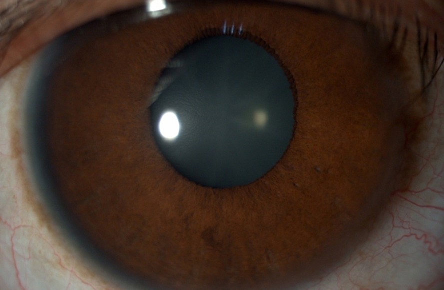
An aphakic eye usually shows iris tremor because no lens supports the iris.
In pseudophakia, a well-centered in-the-bag lens generally keeps the iris quiet.
However, a sulcus lens, an anterior chamber lens or a loose capsular bag may allow movement.
New tremor in a pseudophakic eye should prompt a search for IOL subluxation.
Imaging and Documentation
Ultrasound biomicroscopy shows zonular gaps, lens tilt and ciliary body position.
Anterior segment OCT also measures chamber depth and lens position without contact.
However, neither test replaces careful dynamic observation at the slit lamp.
Therefore, record the sign, its extent and related findings in the preoperative note.
Surgical Implications of Iridodonesis
Preoperative iris tremor warns of zonular weakness before the first incision.
Therefore, the surgeon can plan capsular tension rings, hooks or segments.
A gentle capsulorhexis and careful hydrodissection reduce stress on the remaining zonules.
In addition, low flow settings and a stable chamber limit lens movement during phacoemulsification.
When the Lens Cannot Stay in the Bag
Severe zonular loss may require pars plana lensectomy and vitrectomy.
Then the surgeon chooses between anterior chamber, iris-fixated or scleral-fixated lenses.
Each option carries risks, including endothelial loss, cystoid macular edema and later dislocation.
Discuss this possibility with the patient during consent.
Counseling the Patient With Iridodonesis
Most patients notice nothing, since the tremor itself does not affect vision.
However, explain that the sign suggests weaker lens support.
Tell them that surgery may take longer and may need extra devices.
Also explain that a lens can shift years later, so new blur or edge glare needs prompt review.
Systemic Workup for Iridodonesis
Bilateral tremor with ectopia lentis in a young patient needs a systemic evaluation.
Therefore, consider Marfan syndrome, homocystinuria and Weill-Marchesani syndrome.
Cardiology review and genetic counseling may follow, while homocystinuria also raises thrombotic risk under anesthesia.
Pitfalls to Avoid
- Do not look for the sign only after dilation
- Never assume a quiet iris rules out zonular weakness
- Do not ignore unilateral tremor after old trauma
- Avoid missing pseudoexfoliation on an undilated pupil
- Do not skip the systemic workup in young patients with ectopia lentis
For the latest studies, see the PubMed results for iridodonesis.


Document what you see
Two smartphone imaging tools built for everyday clinic use — one for the slit lamp, one for the fundus.
From Choroida — the team behind this siteReferences
- Schlötzer-Schrehardt U, Naumann GO. Ocular and systemic pseudoexfoliation syndrome. Am J Ophthalmol. 2006;141:921-937.
- Nelson LB, Maumenee IH. Ectopia lentis. Surv Ophthalmol. 1982;27:143-160.
- Hoffman RS, Snyder ME, Devgan U, et al. Management of the subluxated crystalline lens. J Cataract Refract Surg. 2013;39:1904-1915.
- American Academy of Ophthalmology. Basic and Clinical Science Course, Section 11: Lens and Cataract. San Francisco: AAO; 2023.
- Bowling B. Kanski’s Clinical Ophthalmology: A Systematic Approach. 8th ed. Elsevier; 2016.

Writes and reviews the clinical articles on Educate. Every post is checked against peer-reviewed sources before it goes live.