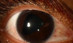
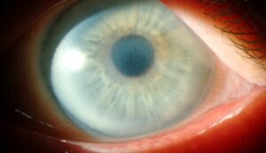
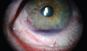
Van Herick test estimates peripheral anterior chamber depth at the slit lamp in under a minute.
It compares the limbal chamber gap with the thickness of the adjacent cornea.
As a result, it flags eyes that need gonioscopy.
However, it screens the angle and cannot replace a direct view of it.
What the Van Herick Test Measures
Van Herick, Shaffer and Schwartz described the method in 1969.
The examiner judges the dark gap between the corneal endothelium and the peripheral iris.
Because this gap lies near the angle recess, it indirectly estimates angle width.
The cornea serves as a built-in ruler.
Slit-Lamp Setup
First, use moderate magnification and a thin, bright slit.
Next, set the illumination arm 60 degrees from the microscope axis.
Then aim the beam perpendicular to the cornea at the temporal limbus.
Finally, dim the room lights so the dark gap stands out (see slit-lamp illumination techniques).
All-fit Slit-Lamp Adapter
Record and share exactly what you see at the slit lamp. One adapter fits any slit lamp or surgical microscope — and any smartphone.
From Choroida — the team behind this siteBeam Position in the Van Herick Test
The slit must fall right at the limbus, just inside clear cornea.
If it lands too far centrally, the chamber looks deeper than it is near the angle.
Meanwhile, the patient looks straight ahead while you view the section from the front.
Grading the Van Herick Test
Compare the dark gap with the corneal section width in the same view.
- Equal to or wider than corneal thickness: grade 4, closure very unlikely
- One quarter to one half: grade 3, closure unlikely
- About one quarter: grade 2, closure possible
- Less than one quarter: grade 1, closure likely
- No visible gap: grade 0 in many modified charts, angle probably closed
The Modified Foster Scale
Foster and colleagues refined the scheme in 2000 for screening in Asian populations.
They recorded limbal chamber depth as a percentage of corneal thickness, using finer steps.
Therefore, the scale helps when you track small changes over time.
How the Van Herick Test Correlates With Gonioscopy
Grades 3 and 4 usually match an open angle on gonioscopy.
However, agreement weakens in the middle grades.
For example, Thomas and colleagues found the test a poor predictor of occludable angles.
As a result, a reassuring grade lowers suspicion without proving the angle is open.
Plateau Iris and Other Exceptions
The test samples one point, usually temporal, while the superior angle is often narrowest.
In plateau iris, the peripheral chamber can look acceptable despite a crowded recess.
Also, peripheral anterior synechiae and angle recession escape detection entirely.
When the Limbus Is Hard to Read
Arcus, pterygium, limbal scars and corneal edema blur the corneal section.
In addition, unusual corneal thickness distorts the ruler, because grading depends on it.
Therefore, record the quadrant you graded and any limbal opacity.
Documenting the Van Herick Test
Write the grade and quadrant for each eye, for example “VH 2 temporal OD.”
Then repeat the test in narrow eyes, because the lens thickens with age.
When to Refer for Gonioscopy
A grade of 2 or lower calls for gonioscopy (see gonioscopy).
Similarly, intermittent halos, brow ache or blur in dim light warrant an angle exam at any grade.
Van Herick Test Before Dilation
Many clinicians run the test before instilling mydriatics.
The risk of dilation triggering acute closure is low, even with narrow angles.
Still, a grade 1 eye should prompt gonioscopy and a talk about warning symptoms.
After dilation, tell patients to seek urgent care for pain, redness, nausea or halos.
When Iridotomy Enters the Discussion
Laser iridotomy decisions rest on gonioscopy, pressure and optic nerve status, never on a Van Herick grade alone.
The ZAP trial found a low rate of progression in untreated angle-closure suspects.
Consequently, routine prophylactic laser now needs individual justification (see angle-closure suspect).
Counseling the Patient
Explain that the test checks how much room the eye’s drainage area has.
A narrow result does not mean glaucoma.
However, it does mean the angle needs a closer look with a contact lens.
Also mention that some medicines, including topiramate, can trigger angle closure.
Van Herick Test Versus Imaging
Anterior segment OCT measures the angle objectively but requires costly equipment.
By contrast, the van Herick test needs only a slit lamp and practice.
Therefore, it stays useful in primary care and outreach screening.
Pitfalls to Avoid
- Do not grade with a wide beam or a narrow arm angle
- Avoid placing the slit inside central cornea
- Never treat grade 3 or 4 as proof of an open angle
- Do not compare nasal and temporal readings
- Never skip gonioscopy in symptomatic eyes
For the latest studies, see the PubMed results for van herick test.


Document what you see
Two smartphone imaging tools built for everyday clinic use — one for the slit lamp, one for the fundus.
From Choroida — the team behind this siteReferences
- Van Herick W, Shaffer RN, Schwartz A. Estimation of width of angle of anterior chamber. Incidence and significance of the narrow angle. Am J Ophthalmol. 1969;68:626-629.
- Foster PJ, Devereux JG, Alsbirk PH, et al. Detection of gonioscopically occludable angles and primary angle closure glaucoma by estimation of limbal chamber depth in Asians: modified grading scheme. Br J Ophthalmol. 2000;84:186-192.
- Thomas R, George T, Braganza A, Muliyil J. The flashlight test and van Herick’s test are poor predictors for occludable angles. Aust N Z J Ophthalmol. 1996;24:251-256.
- He M, Jiang Y, Huang S, et al. Laser peripheral iridotomy for the prevention of angle closure: a single-centre, randomised controlled trial. Lancet. 2019;393:1609-1618.
- Gedde SJ, Chen PP, Muir KW, et al. Primary Angle-Closure Disease Preferred Practice Pattern. Ophthalmology. 2021;128:P30-P70.

Writes and reviews the clinical articles on Educate. Every post is checked against peer-reviewed sources before it goes live.