Corneal sensation testing is a quick bedside check of the trigeminal supply to the ocular surface.
The cornea has one of the densest sensory innervations in the body.
Therefore, reduced feeling often signals nerve damage before the epithelium breaks down.
A careful test takes under a minute and needs simple tools.
Why Corneal Sensation Matters
Corneal nerves drive the blink reflex and reflex tearing.
They also release trophic factors that support epithelial healing.
When sensation falls, the surface dries, heals slowly and may ulcerate without pain.
As a result, patients can present late with a deep defect and little discomfort.
Before You Test Corneal Sensation
First, test before instilling any drop, especially topical anesthetic.
Fluorescein and dilating drops can also blunt the response.
Next, explain the test so the patient does not flinch at a moving object.
Finally, always compare both eyes, because a side difference carries more weight than an absolute value.
All-fit Slit-Lamp Adapter
Record and share exactly what you see at the slit lamp. One adapter fits any slit lamp or surgical microscope — and any smartphone.
From Choroida — the team behind this siteThe Cotton Wisp Method
Twist the tip of a clean cotton bud into a fine wisp.
Ask the patient to look straight ahead, then approach from the temporal side.
This lateral approach keeps the wisp out of view and avoids a menace blink.
Touch the central cornea lightly and note both the blink and the patient’s report.
Then repeat in each quadrant if you suspect sectoral loss.
Dental Floss Testing
Unwaxed dental floss offers a cheap and more uniform alternative.
A fresh strand has a consistent thickness, so successive tests vary less.
However, floss and cotton remain qualitative tools.
They show whether sensation is present, reduced or absent, without measuring degree.
Cochet-Bonnet Esthesiometer
This instrument uses a retractable nylon filament of fixed diameter.
At full length (60 mm) the filament is soft and exerts little pressure.
Shortening it makes the filament stiffer, so it presses harder on the cornea.
The examiner starts long and shortens in steps until the patient feels the touch.
The threshold length then gives a semiquantitative score for follow-up.
Non-Contact and Imaging Methods
Research esthesiometers deliver controlled pulses of air or gas to the cornea.
They can test mechanical, chemical and thermal sensation separately, as Belmonte and colleagues describe.
Meanwhile, in vivo confocal microscopy shows subbasal nerve density and tortuosity.
However, nerve images do not always correlate closely with measured sensation.
Grading Corneal Sensation
Most clinicians record sensation as normal, reduced or absent in each eye.
With the esthesiometer, write the threshold length in millimeters for each zone.
Values close to the maximum length usually indicate normal sensitivity.
A clear drop against the fellow eye should prompt a search for the cause.
Interpreting Corneal Sensation Results
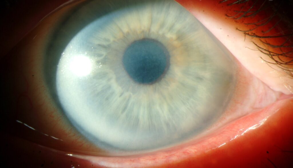
Diffuse bilateral reduction suggests a systemic or chronic surface process.
Sectoral loss fits localized nerve damage, for example after herpes or surgery.
Total unilateral loss raises concern for a lesion along the trigeminal pathway.
Also, correlate the result with staining, tear film and lid closure.
Herpetic Disease and Corneal Sensation
Herpes simplex keratitis often leaves reduced sensation in the affected sector.
Zoster ophthalmicus can cause more profound and widespread loss.
Because of this, a dendrite with poor sensation favors herpetic disease.
Recurrent episodes tend to deepen the deficit over time.
Other Causes of Reduced Corneal Sensation
- Diabetes mellitus with peripheral neuropathy
- Prior LASIK, PRK or penetrating keratoplasty
- Long-term contact lens wear
- Topical anesthetic abuse and chronic preserved drops
- Trigeminal surgery or tumors such as acoustic neuroma
- Some dystrophies, for example lattice corneal dystrophy
- Chemical and thermal burns
Link to Neurotrophic Keratopathy
Neurotrophic keratopathy develops when reduced innervation impairs epithelial maintenance.
Mackie staging moves from punctate keratopathy to persistent epithelial defect, then stromal ulcer.
Dua and colleagues review this progression and its management in detail.
Therefore, test sensation in every nonhealing defect (see stromal corneal ulcers).
Corneal Sensation After Surgery
Refractive surgery cuts subbasal nerves and reduces sensation for months.
Recovery follows nerve regrowth, although it may stay incomplete.
After keratoplasty, graft sensation often returns slowly and partially.
As a result, these eyes need closer surface monitoring.
Counseling the Patient
Explain that a numb cornea may not warn of injury or infection.
Advise prompt review for redness, discharge or blur, even without pain.
In addition, stress regular lubrication and protection from trauma.
An abrasion in a numb eye needs closer follow-up (see corneal abrasion).
Pitfalls to Avoid
- Testing after topical anesthetic or fluorescein
- Approaching from the front and triggering a visual blink
- Testing only the center when herpes suggests sectoral loss
- Recording “normal” without comparing both eyes
- Ignoring total unilateral loss without further assessment
For the latest studies, see the PubMed results for corneal sensation.


Document what you see
Two smartphone imaging tools built for everyday clinic use — one for the slit lamp, one for the fundus.
From Choroida — the team behind this siteReferences
- Cochet P, Bonnet R. L’esthésie cornéenne. Clin Ophthalmol. 1960;4:3-27.
- Müller LJ, Marfurt CF, Kruse F, Tervo TM. Corneal nerves: structure, contents and function. Exp Eye Res. 2003;76:521-542.
- Belmonte C, Acosta MC, Gallar J. Neural basis of sensation in intact and injured corneas. Exp Eye Res. 2004;78:513-525.
- Belmonte C, Nichols JJ, Cox SM, et al. TFOS DEWS II pain and sensation report. Ocul Surf. 2017;15:404-437.
- Dua HS, Said DG, Messmer EM, et al. Neurotrophic keratopathy. Prog Retin Eye Res. 2018;66:107-131.
- Bonini S, Rama P, Olzi D, Lambiase A. Neurotrophic keratitis. Eye (Lond). 2003;17:989-995.
Test yourself
A few questions straight from this article.
-
Besides driving the blink reflex and reflex tearing, how do corneal nerves support the ocular surface?
Corneal nerves release trophic factors for epithelial healing, so when sensation falls the surface dries, heals slowly and may ulcerate without pain. -
When should corneal sensation be tested relative to eye drops?
Testing comes before any drop, especially topical anesthetic, because fluorescein and dilating drops can also blunt the response. -
Why is the cotton wisp brought in from the temporal side when testing corneal sensation?
With the patient looking straight ahead, a lateral approach keeps the wisp out of view, so the blink reflects touch rather than a visual threat. -
What limitation do cotton wisp and dental floss testing of corneal sensation share?
Floss has a more consistent thickness than cotton, but both only show whether sensation is present, reduced or absent. -
How does shortening the Cochet-Bonnet esthesiometer filament change the stimulus?
The examiner starts long and shortens in steps until the touch is felt, because a shorter filament is stiffer and presses harder. -
At what full length is the Cochet-Bonnet esthesiometer filament softest?
At its full 60 mm length the nylon filament is soft and exerts little pressure, and the threshold length gives a semiquantitative score for follow-up. -
What does total unilateral loss of corneal sensation raise concern for?
Total unilateral loss raises concern for a trigeminal pathway lesion, whereas diffuse bilateral reduction suggests a systemic or chronic surface process and sectoral loss fits localized damage. -
Which herpetic disease can cause the more profound and widespread loss of corneal sensation?
Herpes simplex keratitis often leaves reduced sensation in the affected sector, while zoster ophthalmicus can cause more profound and widespread loss. -
What is the order of Mackie staging in neurotrophic keratopathy?
Mackie staging moves from punctate keratopathy to persistent epithelial defect and then stromal ulcer, which is why sensation should be tested in every nonhealing defect. -
Why does refractive surgery reduce corneal sensation for months?
Refractive surgery cuts subbasal nerves, and recovery follows nerve regrowth, although it may stay incomplete, so these eyes need closer surface monitoring.

Writes and reviews the clinical articles on Educate. Every post is checked against peer-reviewed sources before it goes live.