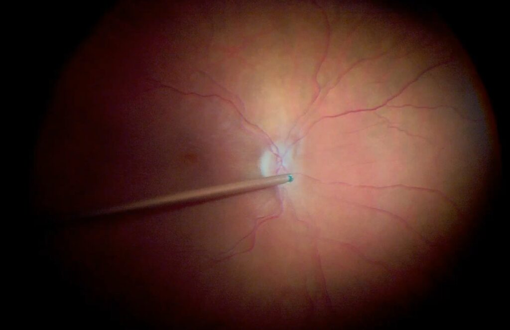
Small-gauge vitrectomy replaced the older 20-gauge technique with narrower instruments that pass through self-sealing transconjunctival ports.
The 23, 25, and 27-gauge systems differ in instrument stiffness, flow rate, and the type of eye they suit.
Knowing these differences helps surgeons choose the right system for each case.
How Small-Gauge Vitrectomy Works
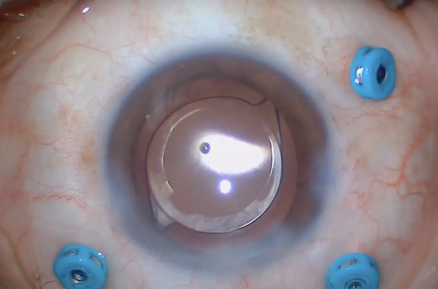
The surgeon places trocar cannulas through the conjunctiva and sclera at an angle, and the wound closes by itself.
The vitreous cutter, light pipe, and infusion line enter through the cannulas.
Compared with 20-gauge surgery, the incisions are smaller, and fewer sutures are needed.
Fujii and colleagues described 25-gauge transconjunctival surgery, and Eckardt later introduced 23-gauge instruments (see pars plana vitrectomy).
Advantages of Small-Gauge Vitrectomy
- Less conjunctival scarring and inflammation
- Shorter operating times for simple cases
- Faster visual recovery and better comfort
- Smaller wounds for repeated surgery
These benefits explain why most vitreoretinal surgeons use small-gauge systems for routine cases.
Fundus Explorer Pro
Photograph the retinal findings described here with the phone already in your pocket — 22 D optics and built-in illumination in one handheld unit.
From Choroida — the team behind this siteChoosing the Gauge
Stiffer 23-gauge instruments allow high flow, so they suit complex detachments and diabetic membrane work.
Meanwhile, 25-gauge instruments balance flexibility and stiffness, and they work well for macular surgery and vitreous hemorrhage.
Fine 27-gauge instruments give access in tight spaces, but they flex more and clear dense vitreous slowly.
Many surgeons mix gauges within a practice, according to the task.
Evidence for Small-Gauge Vitrectomy
Studies show similar anatomic outcomes between 20-gauge and small-gauge vitrectomy for macular surgery and retinal detachment.
Postoperative inflammation is lower, and patients recover faster.
Some series reported higher rates of early hypotony or endophthalmitis with early sutureless wounds, which better port design has reduced (see hypotony maculopathy).
Complications of Small-Gauge Vitrectomy
- Postoperative hypotony from leaking wounds
- Endophthalmitis, with slightly higher risk in early reports
- Vitreous incarceration at the port, with retinal tears
- Instrument flexing, which may reduce control
Suture the port when it leaks, and inspect the wounds at the end of the case.
Tips for Safe Sclerotomies
Displace the conjunctiva before inserting the trocar, so that the wound closes in an offset pattern.
Use an oblique entry, then rotate the cannula to a perpendicular position.
Check each port at the end of surgery, and apply pressure or a suture when leakage persists.
An Example Case
A 63-year-old woman needs surgery for a macular hole.
The surgeon chooses 25-gauge instruments, completes peeling, and fills the eye with gas.
At the first visit, the eye is quiet, the wounds are closed, and she keeps the face-down position for three days.
Key Points
- Choose 23 gauge for stiff, complex work
- Use 25 or 27 gauge for delicate macular surgery
- Always check the wounds at the end
Instruments and Settings
Cutter speed, vacuum, and infusion pressure differ by system, so learn the settings for your platform.
First, use high cut rates near the retina to reduce traction.
Then lower the vacuum when working close to a mobile retina.
Because 27-gauge instruments flow slowly, surgeons often raise the infusion pressure or use a bimanual technique.
Pitfalls to Avoid
- Entering at a perpendicular angle, which makes wounds leak
- Ignoring port leaks at the end of the case
- Using a flexible 27-gauge probe for dense membranes
- Forgetting vitreous incarceration at the trocar sites
Questions Patients Ask
Patients ask whether the surgery needs stitches, and in most cases the ports close without them.
They also ask when they can return to normal life, and many resume light activities within days.
Because recovery depends on the reason for surgery, tailor the advice to the condition.
Looking Ahead
Manufacturers continue to improve cutters and light sources.
Also, higher cut rates and better flow control may let 27-gauge systems handle tougher cases.
However, surgeon skill and case selection still decide the outcome.
Wound Construction in Detail
Start by pulling the conjunctiva to one side with a blunt tool or the cannula itself.
Then insert the trocar at a shallow angle, usually about 30 degrees, and slide it through the sclera.
Next, rotate the cannula to a perpendicular position and push it in until the collar sits on the sclera.
After surgery, remove the cannulas one at a time and press on the wounds with a cotton tip.
Also inspect each wound under the microscope, and place a suture if fluid leaks.
Because early hypotony is the commonest complication, a careful closure pays off.
Finally, record the gauge and any wound sutures in the operative note.


Document what you see
Two smartphone imaging tools built for everyday clinic use — one for the slit lamp, one for the fundus.
From Choroida — the team behind this siteReferences
- Fujii GY, De Juan E Jr, Humayun MS, et al. A new 25-gauge instrument system for transconjunctival sutureless vitrectomy surgery. Ophthalmology. 2002;109:1807-1812.
- Eckardt C. Transconjunctival sutureless 23-gauge vitrectomy. Retina. 2005;25:208-211.
- Oshima Y, Wakabayashi T, Sato T, et al. A 27-gauge instrument system for transconjunctival sutureless microincision vitrectomy surgery. Ophthalmology. 2010;117:93-102.
- American Academy of Ophthalmology. Basic and Clinical Science Course, Section 12: Retina and Vitreous. AAO; current edition.

Writes and reviews the clinical articles on Educate. Every post is checked against peer-reviewed sources before it goes live.