Collateral vessels are new venous channels that form to bypass an occluded retinal vein.
They are a sign of a past or chronic occlusion, and they may help to reduce macular edema.
Recognizing them avoids confusion with neovascularization.
What Collateral Vessels Are
When a retinal vein is blocked, venous pressure rises, and existing capillary connections enlarge.
These vessels form a bypass that drains blood into a nearby unblocked vein.
They are mature, thin-walled, and usually stable, and they do not leak much on angiography (see branch retinal vein occlusion).
Collateral Vessels in Branch Occlusion
In branch retinal vein occlusion, collaterals often cross the horizontal raphe.
They link the occluded territory with the vein on the other side.
Hayreh and Zimmerman described the fundus changes of branch occlusion over time, including these channels.
Fundus Explorer Pro
Photograph the retinal findings described here with the phone already in your pocket — 22 D optics and built-in illumination in one handheld unit.
From Choroida — the team behind this siteOptociliary Shunt Vessels
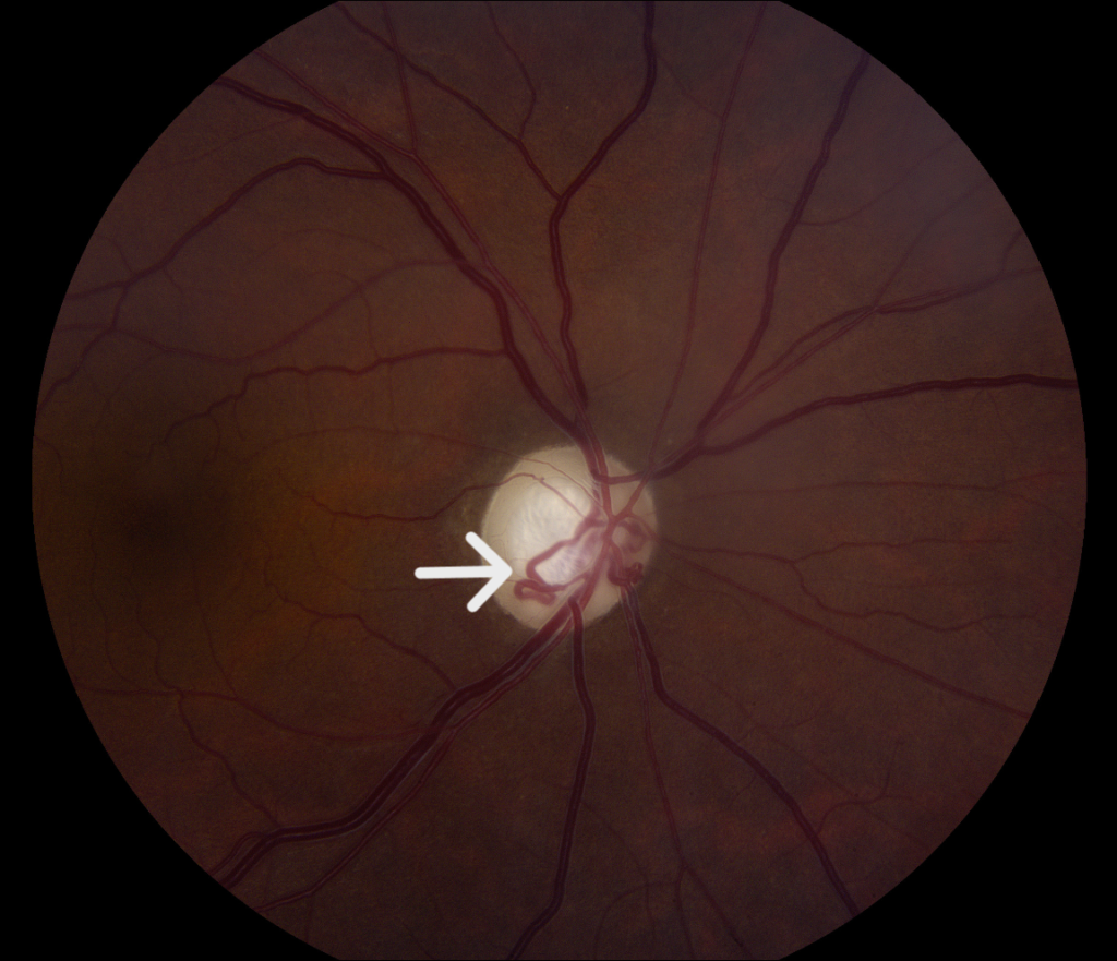
In central retinal vein occlusion, collaterals may appear at the optic disc.
These optociliary shunts link retinal veins to the choroidal circulation at the disc margin.
They also occur with optic nerve sheath meningioma, glaucoma, and chronic papilledema (see central retinal vein occlusion).
How to Recognize Collateral Vessels
- Tortuous, dilated vessels that cross the raphe or run on the disc
- Absence of leakage and fibrous proliferation on angiography
- Location in an area with a history of occlusion and previous hemorrhages
Neovascularization, in contrast, leaks and extends into the vitreous.
What Collateral Vessels Mean
They show that the occlusion is old and that the venous system has adapted.
Hayreh noted that eyes with good collaterals may have less persistent edema.
They do not need treatment by themselves, and laser or injections are not directed at them.
Document them so that later examiners do not mistake them for new vessels.
Practical Points on Collateral Vessels
Use fluorescein angiography when a vessel looks suspicious.
Check for associated ischemia and neovascularization elsewhere.
Reassure patients that the vessels are a natural adaptation.
An Example Case
A 66-year-old man had a branch retinal vein occlusion in the superotemporal arcade two years ago.
His acuity is 20/40, and the exam shows tortuous vessels crossing the horizontal raphe, with a few hard exudates.
Angiography shows collateral channels with no leakage and a region of nonperfusion without neovascularization.
He needs no treatment, and his follow-up is routine.
Questions Patients Ask
Patients ask whether the new vessels are dangerous, and collaterals are a natural bypass, not a growth that damages the eye.
They also ask whether they will block again, and the risk depends on systemic factors that doctors manage.
Because the term new vessels frightens patients, explain the difference.
Pitfalls to Avoid
- Treating collaterals with laser or injections as if they were neovascularization
- Missing true neovascularization at the border of nonperfusion
- Ignoring systemic risk factors such as hypertension and glaucoma
- Failing to document the vessels at baseline
Looking Ahead
OCT angiography shows collaterals clearly without dye and helps to follow them over time.
Also, studies may test whether collateral patterns predict edema and response to treatment.
Therefore, imaging may refine prognosis after vein occlusion.
Practical Points
Use red-free photographs and angiography when the vessels look unusual.
Then compare with older images to confirm stability.
Also review blood pressure, lipids, and glaucoma status in every patient.
How to Tell Collaterals From Neovascularization
Collaterals are tortuous, evenly sized, and do not leak on fluorescein.
Neovascularization appears as fine, frond-like vessels that leak and arise from veins or the disc.
Because the treatment differs, review angiograms carefully.
Systemic Risk Review
Check for hypertension, diabetes, hyperlipidemia, and glaucoma.
Order thrombophilia tests in young patients or those with recurrent events.
Then coordinate care with the primary physician.
Collaterals on OCT Angiography
OCT angiography shows collaterals as large, tortuous vessels in the superficial plexus that connect adjacent venous territories.
Also, it shows the surrounding capillary dropout without the need for dye.
Because the images are quick, they suit follow-up.
Collaterals and Macular Edema
Some patients with collaterals have less persistent edema, because the bypass lowers venous pressure.
However, others keep edema despite well-formed collaterals.
Therefore, treat the edema on its own merits, and do not assume that collaterals protect the macula.
Then follow thickness on OCT at regular intervals.
Summary
Collaterals mark an old occlusion and need no treatment by themselves.
Therefore, document them and look for true neovascularization.
Also continue systemic risk control, since a second occlusion may follow.
For the latest studies, see the PubMed results for collateral vessels.


Document what you see
Two smartphone imaging tools built for everyday clinic use — one for the slit lamp, one for the fundus.
From Choroida — the team behind this siteReferences
- Hayreh SS. Prevalent misconceptions about acute retinal vascular occlusive disorders. Prog Retin Eye Res. 2005;24:493-519.
- Hayreh SS, Zimmerman MB. Fundus changes in branch retinal vein occlusion. Retina. 2015;35:1016-1027.
- Rogers S, McIntosh RL, Cheung N, et al. The prevalence of retinal vein occlusion: pooled data from population studies from the United States, Europe, Asia, and Australia. Ophthalmology. 2010;117:313-319.
- American Academy of Ophthalmology. Basic and Clinical Science Course, Section 12: Retina and Vitreous. AAO; current edition.

Writes and reviews the clinical articles on Educate. Every post is checked against peer-reviewed sources before it goes live.