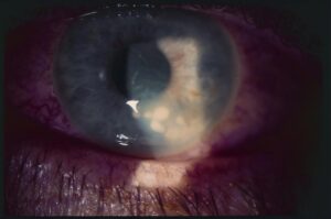
DMEK versus DSAEK is the central comparison in modern endothelial keratoplasty.
Both replace diseased endothelium without a full-thickness graft, but DMEK transplants only a thin layer of Descemet membrane and endothelium, while DSAEK includes a thin layer of stroma (see DSEK and DMEK).
The thinner graft gives better vision and fewer rejections, at the price of a harder operation.
The Two Techniques Compared: DMEK Versus DSAEK
- DSAEK: a graft of endothelium, Descemet membrane, and posterior stroma, about 100 micrometers thick, is placed against the back of the cornea and held with an air bubble
- DMEK: a graft of Descemet membrane and endothelium only, about 15 micrometers thick, is prepared from the donor, scrolled, and unfolded in the eye
DMEK is closer to anatomical replacement.
DMEK Versus DSAEK: Visual Outcomes
In prospective series, a large majority of DMEK eyes reached 20/25 or better.
A significant share reached 20/20, which is rarer with DSAEK (see the references).
The stromal interface in DSAEK adds some light scatter and a hyperopic shift, and many DSAEK eyes plateau around 20/30 to 20/40.
Visual recovery is also faster with DMEK, often within weeks.
All-fit Slit-Lamp Adapter
Record and share exactly what you see at the slit lamp. One adapter fits any slit lamp or surgical microscope — and any smartphone.
From Choroida — the team behind this siteRejection and Graft Survival
- Rejection rates after DMEK are very low, around 1 percent in many series
- Rates after DSAEK are higher, often several percent to about 10 percent, and penetrating keratoplasty has the highest rates (see the references)
- Less tissue means less antigen, so steroid use after DMEK can be shorter and lower in dose, with less risk of pressure rise (see steroid-induced glaucoma)
Complications of DMEK Versus DSAEK
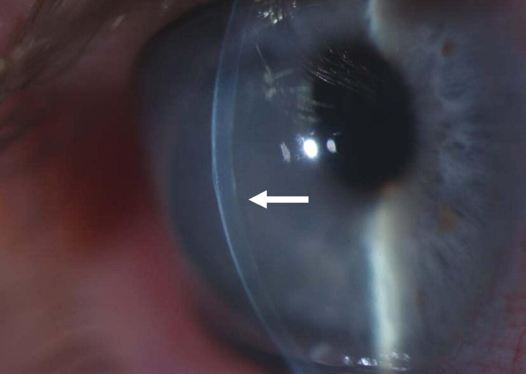
- Graft detachment: more common with DMEK, because the thin graft attaches by air alone, and rebubbling is needed in a fair proportion of eyes
- Primary graft failure: low in both, with a slightly higher rate in DMEK in early series
- Endothelial cell loss: similar over time
- Interface haze and thick graft problems: unique to DSAEK
DMEK Versus DSAEK: Learning Curve and Tissue
DMEK demands more skill, a longer learning period, and careful donor tissue handling, since the graft can tear during preparation.
Eye banks now prepare many DMEK grafts, which saves time and tissue (see eye banking and the donor cornea).
DSAEK is easier to learn and more forgiving, and works in eyes with unusual anatomy.
Choosing the Technique
- DMEK: the usual choice for Fuchs dystrophy and pseudophakic edema with a normal anterior chamber
- DSAEK: useful in eyes with difficult anatomy, such as aphakia, a missing iris, an anterior chamber lens, or a previous glaucoma tube, where graft retention is difficult
- Penetrating keratoplasty: for scarred cornea (see penetrating keratoplasty)
Follow-Up for DMEK Versus DSAEK
- Review the first day for bubble position and graft attachment
- Check again at one week and one month, with OCT in unclear cases
- Taper steroids over months, and monitor the pressure
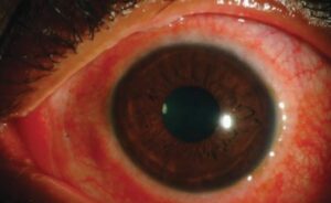
- Watch for rejection signs, such as redness, pain, or blur
Counselling
Explain that most patients see well after either method, that DMEK gives the best vision and lowest rejection, and that a second bubble may be needed.
An Example Case
A 68-year-old woman with Fuchs dystrophy and pseudophakic eyes has corneal edema and 20/80 vision.
She receives a DMEK graft, with an air bubble, and lies flat for a day or two, and by six weeks she sees 20/25, with a clear cornea (see Fuchs endothelial corneal dystrophy).
A small detachment at the edge needed a second bubble, which resolved the issue in a day.
Preoperative Planning
- Review the anterior chamber depth, iris status, and any previous glaucoma surgery
- Choose pre-stripped or surgeon-prepared tissue, according to the supply and experience of the team
- Plan the position and lens status, and discuss the chance of rebubbling
- Check the pressure, since glaucoma may complicate management
Long-Term Care
Most grafts remain clear for many years.
Patients should keep up with checks of pressure, graft clarity, and endothelial counts when needed.
Rejection episodes are usually reversible if treated early with steroids (see corneal graft rejection).
Training Tips
Surgeons new to DMEK can start with simple cases, such as pseudophakic Fuchs dystrophy with a clear view, and use pre-prepared tissue.
Practice on eye bank training tissue helps to learn how to unfold the graft with minimal handling.
Key Messages for Colleagues
Choose the technique by anatomy and experience, and offer DMEK when it is feasible, since the results are better.
Train for DMEK in steps, plan for rebubbling, and keep good records of outcomes, including rejection and detachment rates.


Document what you see
Two smartphone imaging tools built for everyday clinic use — one for the slit lamp, one for the fundus.
From Choroida — the team behind this siteReferences
- Price MO, Giebel AW, Fairchild KM, Price FW Jr. Descemet’s membrane endothelial keratoplasty: prospective multicenter study of visual and refractive outcomes and endothelial survival. Ophthalmology. 2009;116:2361-2368.
- Guerra FP, Anshu A, Price MO, Giebel AW, Price FW. Descemet’s membrane endothelial keratoplasty: prospective study of 1-year visual outcomes, graft survival, and endothelial cell loss. Ophthalmology. 2011;118:2368-2373.
- Hos D, Matthaei M, Bock F, et al. Immune reactions after modern lamellar (DALK, DSAEK, DMEK) versus conventional penetrating corneal transplantation. Prog Retin Eye Res. 2019;73:100768.
- American Academy of Ophthalmology. Basic and Clinical Science Course, Section 8: External Disease and Cornea. AAO; current edition.

Writes and reviews the clinical articles on Educate. Every post is checked against peer-reviewed sources before it goes live.