Diabetic keratopathy is a group of corneal changes caused by diabetes, in which the nerves, the epithelium, and the tear film are all affected.
It is often overlooked, since patients may have few symptoms, but it becomes obvious when surgery or injury stresses the cornea.
In addition, recognizing it protects the eye from slow healing and recurrent defects.
What Is Diabetic Keratopathy?
- Neuropathy: loss of corneal nerve fibers, with lower corneal sensation, found on confocal microscopy and esthesiometry (see the references)
- Epithelial changes: fragile cells, thick basement membrane, and poor adhesion
- Tear film problems: reduced tear production and an unstable film, with dry eye
- Slow healing and reduced wound response
- Endothelial changes, with polymegethism, and a lower ability to recover from stress
Poor glycemic control worsens these features, and they are more marked in patients with retinopathy and peripheral neuropathy (see the references).
Diabetic Keratopathy: Clinical Presentation
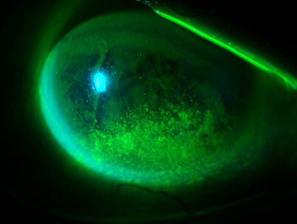
- Punctate keratopathy and superficial erosions
- Recurrent corneal erosions
- Persistent epithelial defects, especially after surgery (see persistent epithelial defect)
- Corneal ulcers, with a higher risk of infection and a slower response
- Reduced blink and a quiet, painless eye, since sensation is low
Many patients have no symptoms until a defect appears.
All-fit Slit-Lamp Adapter
Record and share exactly what you see at the slit lamp. One adapter fits any slit lamp or surgical microscope — and any smartphone.
From Choroida — the team behind this siteWhen It Becomes a Problem
- After vitrectomy, when the epithelium is removed to improve the view, and may fail to regrow for weeks
- After cataract surgery and laser, with prolonged healing and a higher risk of NSAID melt (see NSAID keratopathy)
- Following corneal transplantation, with poor epithelial healing and neurotrophic ulcers
- During contact lens wear, with a higher risk of infection
- In patients on topical medications, such as preserved glaucoma drops
Diabetic Keratopathy: Assessment
- Check the corneal sensation, with a cotton wisp or esthesiometer (see corneal sensation testing)
- Then look for punctate staining and the quality of the tear film
- Examine the epithelium for irregularity and map-like patterns
- Review the control of diabetes, and the other complications
- Consider imaging of corneal nerves, where available
Management for Diabetic Keratopathy
- Optimize glycemic control, with the diabetes team
- Lubricants and lid care, with preservative-free formulas
- Avoid toxic drops, and review all medications
- Protect the epithelium during surgery: minimize epithelial debridement, use intraoperative lubrication, and consider alternatives to removal in vitrectomy
- Treat defects early, with a bandage lens, serum drops, or amniotic membrane (see autologous serum eye drops)
- Nerve growth factor for neurotrophic ulcers, where it is available
- Close follow-up after any surgery
Aldose reductase inhibitors and other agents have been tested, but results are limited.
Practical Advice for Surgeons
Before intraocular surgery in a diabetic patient, assess sensation and the ocular surface, and warn the patient about slow healing.
Also, use short courses of NSAID, and look at the cornea at each visit.
Document the findings, and plan for early intervention if an epithelial defect appears.
Counselling
Explain that the cornea of a person with diabetes is more fragile, and that the eye may not hurt even when it is damaged.
Teach the patient to report redness, discharge, or blurred vision at once, and to attend all follow-up visits.
A Typical Case
A 58-year-old man with type 2 diabetes for twenty years and proliferative retinopathy has vitrectomy with epithelial debridement for a better view.
Four weeks later, a central epithelial defect remains, with reduced sensation and no pain.
The team stops preserved drops and starts autologous serum and a bandage lens, and the defect heals over three weeks.
The doctor advises him that the cornea will remain fragile, and that his glaucoma and dry eye drops should be preservative-free (see dry eye disease).
Diabetic Keratopathy and Infection
Diabetic patients have a higher risk of corneal infection after contact lens wear and surgery, and ulcers heal slowly.
Also, culture early in any infiltrate, treat intensively, and review often.
Monitoring in Clinic
- Check corneal sensation at least yearly in patients with neuropathy
- Look at the epithelium at every visit, and ask about dry eye symptoms
- Encourage regular eye examinations for retinopathy, since examiners find many corneal problems at the same time
Research Directions
Studies are exploring nerve growth factor, substance P, and other agents that could improve healing and nerve recovery.
Researchers are studying corneal confocal microscopy as a marker of diabetic neuropathy elsewhere in the body.
Patient Education
Show patients how to inspect their eyes for redness, and remind them that a painless red eye still needs prompt attention.
Also, provide a list of preservative-free options for their regular drops, and ask them to bring all their medications to each visit.
For the latest studies, see the PubMed results for diabetic keratopathy.


Document what you see
Two smartphone imaging tools built for everyday clinic use — one for the slit lamp, one for the fundus.
From Choroida — the team behind this siteReferences
- Schultz RO, Van Horn DL, Peters MA, Klewin KM, Schutten WH. Diabetic keratopathy. Trans Am Ophthalmol Soc. 1981;79:180-199.
- Bikbova G, Oshitari T, Baba T, Bikbov M, Yamamoto S. Diabetic corneal neuropathy: clinical perspectives. Clin Ophthalmol. 2018;12:981-987.
- Han SB, Yang HK, Hyon JY. Influence of diabetes mellitus on anterior segment of the eye. Clin Interv Aging. 2019;14:53-63.
- American Academy of Ophthalmology. Basic and Clinical Science Course, Section 8: External Disease and Cornea. AAO; current edition.

Writes and reviews the clinical articles on Educate. Every post is checked against peer-reviewed sources before it goes live.