
A tigroid fundus, also called a tessellated fundus, describes a striking striped or tiger-skin appearance visible on dilated fundus examination, and recognizing what actually produces this pattern, thinning of the retinal pigment epithelium unmasking the underlying choroidal blood vessels, explains both why it looks the way it does and why its presence carries real information about the health and risk profile of the eye it is found in.
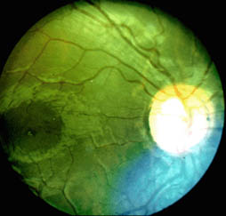
What Produces the Striped Appearance
The retinal pigment epithelium normally contains enough pigment to substantially obscure the underlying choroidal vasculature from view during fundus examination, giving the normal fundus its relatively uniform, orange-red appearance.
When the retinal pigment epithelium thins, as commonly occurs with axial elongation in myopia, this pigment layer becomes less effective at masking the choroid beneath it, and the large choroidal vessels become directly visible as alternating light and dark stripes, producing the characteristic tigroid or tessellated pattern.
Myopia as the Dominant Cause
A tigroid fundus is most commonly seen in myopic eyes, and its prevalence and prominence generally increase with the degree of myopia and, to some extent, with age, reflecting the progressive retinal pigment epithelial thinning that accompanies axial elongation of the eye over time.
- Mild to moderate tigroid change is common and often not, by itself, a marker of significant pathology
- More pronounced tessellation, particularly in high myopia, is more often associated with the broader spectrum of myopic fundus changes and should prompt a more thorough assessment for other signs of pathologic myopia
Fundus Explorer Pro
Photograph the retinal findings described here with the phone already in your pocket — 22 D optics and built-in illumination in one handheld unit.
From Choroida — the team behind this siteWhat a Tigroid Fundus Signals in High Myopia
In the context of high myopia, a prominent tigroid appearance is often one of the earliest visible fundus changes reflecting the underlying process of progressive chorioretinal thinning, and its presence should prompt a careful, systematic search for other, more directly vision-threatening features of pathologic myopia.
- Posterior staphyloma, a structural outward bulging of the thinned sclera and overlying tissue (see posterior staphyloma)
- Lacquer cracks, breaks in Bruch membrane that can develop within areas of significant chorioretinal thinning
- Areas of more focal or diffuse chorioretinal atrophy, which can progress over time and, in advanced cases, threaten central vision if they extend into or near the macula
- Increased risk of myopic choroidal neovascularization, related to the same underlying thinning and structural change
Clinical Evaluation
- Dilated fundus examination characterizing the extent and distribution of the tessellated pattern, along with a systematic search for any coexisting signs of more advanced myopic degeneration
- Measurement of axial length and refractive error, since the degree of myopia correlates with both the prominence of tessellation and the broader risk of associated pathologic changes
- Wide-field fundus photography supports documentation and comparison over time, useful for monitoring any progression of associated chorioretinal changes
- Optical coherence tomography can help assess retinal pigment epithelial and choroidal thickness directly, and screen for any early signs of the more serious complications associated with high myopia
Distinguishing a Simple Tigroid Fundus From Pathologic Myopia
The key clinical distinction is between a tigroid appearance as an isolated, relatively benign finding, common in mild to moderate myopia, and tessellation occurring as one component of a broader pattern of pathologic myopic degeneration, which carries meaningfully different implications for monitoring and long-term visual prognosis.
This distinction is made by assessing the fundus as a whole, specifically looking beyond the tessellation itself for the other structural changes, staphyloma, chorioretinal atrophy, and lacquer cracks, that together define more advanced myopic maculopathy.
Management
A tigroid fundus itself requires no treatment, since it is a descriptive finding reflecting underlying retinal pigment epithelial thinning rather than a disease process in its own right.
When it occurs in the context of high myopia, management is directed at appropriate monitoring for the associated complications of pathologic myopia, including regular dilated examination and patient education about symptoms, such as new distortion or a scotoma, that would warrant prompt reevaluation for a developing complication like choroidal neovascularization.
Prognosis
An isolated tigroid fundus in a patient with mild to moderate myopia carries an excellent prognosis and generally needs nothing beyond routine, age-appropriate eye care.
When tessellation is prominent and accompanies other signs of high, pathologic myopia, the prognosis is tied to that broader condition, and ongoing monitoring for progressive chorioretinal atrophy and neovascular complications becomes the central, appropriately more vigilant, focus of long-term care.


Document what you see
Two smartphone imaging tools built for everyday clinic use — one for the slit lamp, one for the fundus.
From Choroida — the team behind this siteReferences
- Ohno-Matsui K, Kawasaki R, Jonas JB, et al. International photographic classification and grading system for myopic maculopathy. Am J Ophthalmol. 2015;159:877-883.
- Hayashi K, Ohno-Matsui K, Shimada N, et al. Long-term pattern of progression of myopic maculopathy: a natural history study. Ophthalmology. 2010;117:1595-1611.
- Fang Y, Yokoi T, Nagaoka N, et al. Progression of myopic maculopathy during 18-year follow-up. Ophthalmology. 2018;125:863-877.
- Grossniklaus HE, Green WR. Pathologic findings in pathologic myopia. Retina. 1992;12:127-133.
Test yourself
A few questions straight from this article.
-
What produces the striped appearance of a tigroid fundus?
When the retinal pigment epithelium thins it no longer masks the choroid, and the large choroidal vessels become visible as alternating light and dark stripes. -
In which refractive state is a tigroid fundus most commonly found?
A tigroid fundus is most commonly seen in myopic eyes, and its prevalence and prominence generally increase with the degree of myopia and, to some extent, with age. -
What treatment does a tigroid fundus itself require?
A tigroid fundus needs no treatment because it is a descriptive finding reflecting underlying retinal pigment epithelial thinning rather than a disease in its own right. -
In high myopia, what does a prominent tigroid appearance usually represent?
Prominent tessellation in high myopia is often one of the earliest visible fundus changes of progressive chorioretinal thinning, prompting a search for other pathologic myopic features. -
Lacquer cracks seen in pathologic myopia represent breaks in which structure?
Lacquer cracks are breaks in Bruch membrane that can develop within areas of significant chorioretinal thinning. -
A posterior staphyloma in pathologic myopia is best described as what?
Posterior staphyloma is a structural outward bulging of the thinned sclera and the overlying tissue, and is one of the features sought in pathologic myopia. -
Which measurements are taken to gauge risk in an eye with a tessellated fundus?
Axial length and refractive error are measured because the degree of myopia correlates with both the prominence of tessellation and the risk of associated pathologic change. -
Which investigation assesses retinal pigment epithelial and choroidal thickness directly in a tigroid fundus?
Optical coherence tomography assesses retinal pigment epithelial and choroidal thickness directly and screens for early signs of the serious complications of high myopia. -
Which new symptoms should a highly myopic patient with a tigroid fundus be told to report promptly?
Patients are educated to report new distortion or a scotoma promptly, since these suggest a developing complication such as choroidal neovascularisation. -
What is the prognosis of an isolated tigroid fundus in mild to moderate myopia?
An isolated tigroid fundus in mild to moderate myopia carries an excellent prognosis and generally needs nothing beyond routine, age-appropriate eye care.
