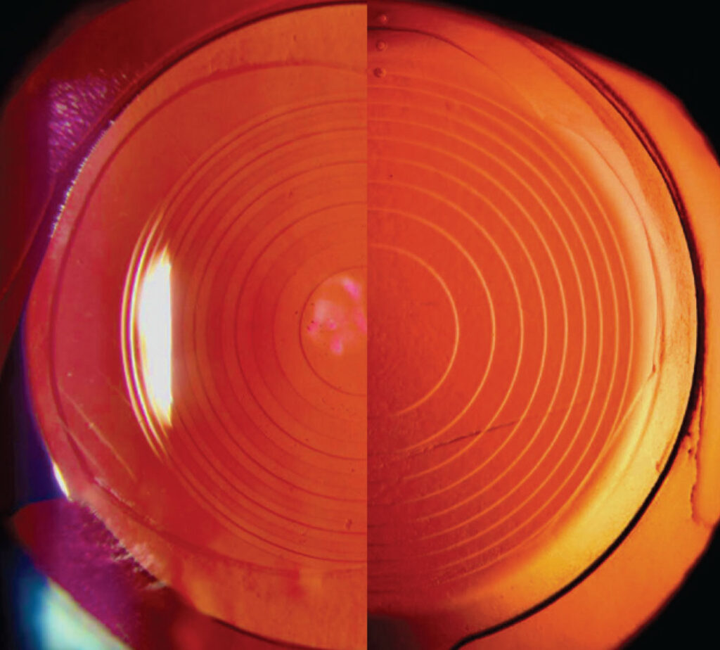
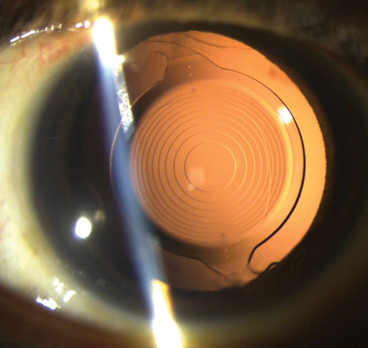
An IOL exchange is the removal of an implanted intraocular lens and its replacement with another.
It is not a common operation, but it solves problems that glasses and drops cannot, such as an unacceptable refractive error or a lens that has failed.
Timing and planning decide how hard it is.
Why an IOL Exchange Is Done
- Dysphotopsia and poor tolerance of multifocal lenses, which is one of the most frequent reasons in recent series (see the references)
- Refractive surprise, from a wrong power, wrong lens, or biometry error
- Dislocation or subluxation, from weak zonules or capsular problems (see dislocated intraocular lens)
- Opacification of the lens, such as calcification of some hydrophilic acrylic lenses
- Uveitis-glaucoma-hyphema syndrome from chafing of the iris or ciliary body (see uveitis-glaucoma-hyphema syndrome)
- Toxic reactions and other complications, including significant glistenings or optic damage
Before exchanging a lens, consider other solutions, such as neuroadaptation, laser, or a piggyback lens.
IOL Exchange: Timing
- Early exchange, within weeks to a few months: the capsular bag has not fused with the lens, so the lens is easier to remove and replace
- Late exchange: the capsule is fibrotic, haptics are embedded, and the procedure is harder and has more risk to the zonules and capsule
Patients with a clear indication should not wait, though the first few weeks allow the refraction and the symptoms to settle.
All-fit Slit-Lamp Adapter
Record and share exactly what you see at the slit lamp. One adapter fits any slit lamp or surgical microscope — and any smartphone.
From Choroida — the team behind this sitePlanning
- Review the original operative notes and the type of lens, and its haptic design
- Measure the eye again, and decide the target and the new lens power
- Examine the cornea, the pupil, and the zonules, and the endothelial cell count
- Plan for the replacement lens, which may go in the bag, the sulcus, or the sclera (see scleral-fixed lens)
- Prepare for possible vitrectomy
- Discuss the risks, including those of vitreous loss and macular edema
IOL Exchange: Surgical Technique
- Fill the chamber with a cohesive viscoelastic, and separate the optic and haptics from the capsule with a spatula and viscoelastic
- Rotate the lens into the anterior chamber and cut it into pieces with scissors or a cutter, for removal through a small incision, or remove it whole through a larger one
- Preserve the capsular bag, and check its integrity
- Implant the new lens in the bag if the bag is intact, or in the sulcus with a three-piece lens and optic capture when the bag is damaged
- If there is no capsular support, use a sutured, glued, or iris-fixated lens, or an anterior chamber lens
Take care with the endothelium, since the cornea suffers during manipulation of the lens.
Outcomes and Risks of IOL Exchange
Most patients improve in symptoms and in vision after a well-planned exchange, particularly for multifocal intolerance, though some remain unhappy because of other causes, such as dry eye.
Also, risks include zonular dialysis, vitreous loss, macular edema, endothelial loss, glaucoma, and retinal detachment (see the references).
Counselling
Explain the reasons, the expected benefit, and the uncertainty.
Reassure patients that a second surgery can improve the problem in most cases, and explain the recovery.
An Example Case
A patient had a trifocal lens implanted six weeks ago and complains of severe night halos and blurred vision, despite a normal examination and a correct refraction.
After adaptation, dry eye treatment, and a trial of drops that constrict the pupil, the symptoms persist.
The patient chooses an exchange to a monofocal lens.
At surgery, the lens is easily separated from the bag, since little time has passed.
The surgeon places a monofocal lens in the bag with a target near emmetropia.
The symptoms resolve within days, and the patient is satisfied.
Choosing the Replacement Lens
- Clinicians choose a monofocal lens most often when the problem is dysphotopsia
- A toric or other specific lens may be required when the cause is astigmatism
- Clinicians prefer hydrophobic acrylic lenses in many cases, because of their good clarity and low risk of calcification
- The power is based on new biometry and on the knowledge of the actual position of the old lens
A refractive surprise after an exchange is possible, so the plan should include a discussion of options such as laser correction.
Avoiding the Need for Exchange
- Careful selection of patients for premium lenses
- Treating the ocular surface before surgery
- Accurate biometry, with formulas suited to the eye
- A frank discussion of expectations and of the neuroadaptation period
These steps could avoid many cases of exchange, as could giving the eye time to settle.
For the latest studies, see the PubMed results for IOL exchange.


Document what you see
Two smartphone imaging tools built for everyday clinic use — one for the slit lamp, one for the fundus.
From Choroida — the team behind this siteReferences
- Jones JJ, Jones YJ, Jin GJC. Indications and outcomes of intraocular lens exchange during a recent 5-year period. Am J Ophthalmol. 2014;157:154-162.
- Mamalis N, Brubaker J, Davis D, Espandar L, Werner L. Complications of foldable intraocular lenses requiring explantation or secondary intervention: 2007 survey update. J Cataract Refract Surg. 2008;34:1584-1591.
- Kohnen T, Koch DD, eds. Cataract and Refractive Surgery. Springer; 2005.
- American Academy of Ophthalmology. Basic and Clinical Science Course, Section 11: Lens and Cataract. AAO; current edition.

Writes and reviews the clinical articles on Educate. Every post is checked against peer-reviewed sources before it goes live.